Cardiologia Croatica
Istraživački asistent časopisa
Istraživački asistent časopisa
Istraživački asistent časopisa
Istraživački asistent časopisa
Irzal Hadžibegović, Nikola Pavlović, Tomislav Šipić, Marin Pavlov, Ivana Jurin, Aleksandar Blivajs, Matija Vrbanić, Ante Lisičić, Luka Antolković, Šime Manola
**Introduction**: Functional coronary testing rather than systematic stenting in chronic coronary syndromes has been advocated by the most recent guidelines. However, its use in patients with acute coronary syndromes (ACS) is controversial. (1) We aimed to evaluate the penetration of functional coronary testing in patients with ACS in our center. **Patients and Methods**: We analyzed data from quantitative coronary angiography and functional coronary testing in patients who underwent invasive coronary angiography in 2022. **Results**: Out of 2075 patients who underwent coronary angiography in 2022, 536 (26%) patients had acute coronary syndrome. Among 536 acute coronary syndrome patients, 42 (8%) patients received invasive functional testing. All patients had non-hyperemic indices (instantaneous wave-free ratio [iFR] or coronary resistance reserve [cRR]) analyzed, with 4 (10%) patients with borderline results requiring additional fractional flow reserve (FFR). In 40/42 patients functional testing was done in another setting, in order to assess non-culprit lesions. In only 2 patients functional testing was done during invasive assessment of an acute coronary episode (1 in the circumflex artery territory, and 1 in the proximal left anterior descending artery) and in both cases percutaneous coronary intervention (PCI) was deferred. Generally, there were no adverse events in deferred patients during 6-months follow-up. **Conclusion**: Functional testing in patients with acute coronary syndromes was mostly performed in a staged procedure in order to assess non-culprit lesions. The use of functional testing to defer or confirm PCI indication in a suspected culprit lesion in acute setting, or ad hoc in by-stander lesions immediately after culprit lesion PCI, is still controversial and requires additional study. Additional imaging methods would probably help identify culprit lesions among intermediate stenoses found in patients with acute coronary syndromes.
Lea Saftić, Nikica Prpić, Ivan Gorički, Marko Gatarić
A giant aneurysm is a restricted expansion of a blood vessel. The most common aneurysms are found on the aorta, the largest blood vessel in the body. A giant aneurysm can also be found on coronary arteries and, thus, on peripheral blood vessels. Giant aneurysms of coronary arteries are rare, accounting for 0.02% - 0.2%. The cause of giant aneurysms is a deterioration of the blood vessel wall. Other causes of aneurysms may include atherosclerosis, congenital diseases, Kawasaki disease in children, Takayasu’s arteritis, connective tissue disease, vasculitis, and coronary artery trauma, as a result of percutaneous coronary intervention. Clinical outcomes of giant aneurysms may include thrombus formation, rupture, embolization, and fistula formation. Resections of giant coronary aneurysms can be performed surgically and by percutaneous invasive approach. (1, 2) This paper is a case report on a patient diagnosed with a giant aneurysm of the left coronary artery (50x45x40mm). This paper presents an invasive percutaneous coronary intervention which successfully excluded the giant aneurysm from the coronary circulation.
Martina Čančarević, Mislav Nedić, Kristijan Đula, Siniša Car, Diana Delić-Brkljačić, Vjekoslav Radeljić
The most common symptom of ischemic heart disease is angina pectoris. Ischemia with non-obstructive coronary artery disease (INOCA) is defined as presence of signs and symptoms of ischemia and coronary artery stenosis less than 50% on invasive coronary angiography. INOCA endotypes are microvascular angina (MVA), vasospastic angina (VSA) and mixed MVA and VSA. INOCA patients are more likely to be female aged 40 to 70 years. Smoking is a risk factor for vasospastic angina, while diabetes and hypertension are not (1). The prognosis of patients with INOCA is not benign. Symptoms are often disabling, and patients have higher incidence of adverse cardiovascular events. Treatment of INOCA patients includes lifestyle modifications, management of common risk factors and antianginal medication. Acetylcholine is parasympathetic nervous system neurotransmitter. The ACh spasm provocation test has become a popular method for induction of coronary spasm. Due to rapid degradation by acetylcholinesterase ACh-provocation coronary spasm is short of duration. ACh is injected into coronary artery within 20 seconds with continuous monitoring of the ECG and patient’s symptoms. ENCORE study protocol proposes incremental doses of 2, 20, 100, and 200 micrograms of ACh manually infused into the left coronary artery (LCA). In patients who remain asymptomatic 80 micrograms of ACh is injected into the right coronary artery. Bradycardia and atrio-ventricular block are often during test, but short of duration and usually resolve spontaneously within few minutes (2). Results are based on three criteria. Symptoms such as chest pain or shortness of breath. Second are ECG changes such as ST segment elevation, depression, or T wave changes and third criteria are focal or defuse epicardial diameter reduction of 90% or more. If only first two criteria are met than microvascular angina is diagnosed. Serious complications such as myocardial infarction or ventricular tachycardia/fibrillation are reported in 0.3-1% of cases (3). In conclusion, INOCA patients are often under-diagnosed, under-treated and because of that have worse prognosis. ACh spasm provocation should be performed in patient with specific anginal symptoms, evidence of ischemia and non-obstructive coronary artery disease to guide optimal treatment and improve quality of life.
Tomislav Šipić, Ivana Jurin, Nikola Pavlović, Marin Pavlov, Aleksandar Blivajs, Šime Manola, Irzal Hadžibegović
**Introduction**: Highly calcified coronary lesions represent up to 30% of all coronary stenoses, and bring greater risk of complications and recurrent events. Non-compliant, cutting/scoring and super-high pressure balloons, together with debulking devices (rotablation and intravascular lithotripsy) are essential in calcium management. (1) **Patients and Methods**: We aimed to investigate changes in patterns of calcium management in a tertiary institution cath-lab from 2018 to 2022. We compared yearly penetration of calcium debulking techniques in one center from 2018 to 2022 among all percutaneous coronary intervention (PCI) in patients with chronic and acute coronary syndromes. **Results**: Among all patients treated in 2018 and 2019, rotablation was used in 1.7% and 1.5% of all PCI cases, whereas super-high pressure balloons were used in 1.3% and 1.1% of cases for debulking. Non-compliant balloons were used for lesion preparation in 18% of all PCI. In 2021 and 2022, rotablation was used for debulking in only 0.4% of cases, whereas super-high pressure balloons were used in 1.2% of cases, often in combination with rotablation. Also, in 2021 and 2022 intravascular lithotripsy (IVL) was used in 1.4% of all PCI cases for debulking, and non-compliant balloons were used for lesion preparation in 32% of cases. Among all PCI in 2021 and 2022, there was 1 case of failed stent delivery in a tortuous calcified vessel, and 1 case of inadequate stent expansion after IVL that was successfully expanded with a super-high pressure balloon. **Conclusion**: In cases of highly calcified coronary lesions, IVL has been becoming a standard in crossable lesions, whereas rotablation has been used occasionally - almost exclusively in non-crossable lesions in combination with super-high pressure balloons after successful crossing. Also, routine use of non-compliant ballons for lesion preparation has increased markedly, whereas scoring balloons were not used routinely in calcified lesions.
Hrvoje Lukić, Tomislav Biloglav, Željka Roginić
**Introduction**: Transcatheter aortic valve implantation (TAVI) is a method for the treatment of patients with severe aortic stenosis (AS). These patients are mostly older people with a lot of comorbidities and a high risk for cardiac surgery. TAVI approach includes transfemoral, transaortic, transapical, trans-subclavian, and transcarotid. As with any other invasive procedure, TAVI carries the risk of complications. Perioperative complications include bleeding, valvular complications, arrhythmias, coronary artery occlusion, cerebrovascular insult, left ventricular perforation, and death. Postoperative complications include paravalvular regurgitation, valve thrombosis, bleeding, endocarditis, and death. (1) Aim: To present the complications of transcatheter aortic valve implantation (TAVI) and their management. **Case report**: 84-year-old patient with symptomatic severe AS, comorbidities, and high risk for cardiac surgery. The valvular heart team has recommended TAVI implantation. The TAVI procedure was performed through a transfemoral approach after arterial puncture and placement of sheaths in both the right and left femoral arteries. After placing the wire in the left ventricle, the aortic valve was predilated with a 24 mm balloon, followed by the implantation of the L-size valve. Unfortunately, the valve migrated to the aortic root when the “delivery” system was pulled out. The valve was extracted using the SNARE system and a biopsy (forceps) and positioned in the ascending aorta. Then a new valve was placed with the optimal position that was confirmed by aortography. During the closure of the right femoral artery with the Manta system inadequate anchoring occurred due to the calcified plaque, which resulted in subocclusion of the artery shown on control angiographically. Therefore, the “cross-over” technique was used to approach the lesion from the left side to perform predilatation and implantation of the stent graft with an optimal final angiographic result. **Conclusion**: TAVI is a minimally invasive treatment method used in patients with severe AS, comorbidities, and high risk for surgery. The most common complications in TAVI patients include vascular complications related to femoral access and paravalvular regurgitation.
Petra Radić, Vjekoslav Radeljić, Matias Trbušić, Mislav Nedić
**Introduction**: Takotsubo syndrome (TTS) was first described in Japan in 1991 as a syndrome affecting predominantly postmenopausal women after emotional stress (1). TTS includes chest pain, ECG changes and wall motion abnormalities as well as elevation of the cardioselective enzymes, which also corresponds to acute myocardial infarction (AMI). Although the etiology of TTS has not yet been clarified, catecholamine-mediated cardiotoxicity provoked by emotional or physical stress is considered one of the most likely causes (2). **Case report**: 65-year-old female was examined in the Emergency Department because of chest pain lasting several hours, which was provoked by a stressful event. The patient stated that she performed cardiopulmonary resuscitation a day earlier on her husband, who suffered a heart attack. In the electrocardiogram on admission, inferolateral ST-segment depression with elevation in AVR was recorded. Echocardiography showed hypokinesia of the middle and apical segment of the inferoposterior wall and ejection fraction of the left ventricle was 55%. An emergency coronary angiography was performed, which showed the occlusion of the circumflex artery (LCx) in the proximal segment. She underwent percutaneous coronary intervention (PCI) with successful stent placement in the LCx. Due to the “slow flow” phenomenon, eptifibatide was administered. During the procedure, the patient developed pulmonary edema and was intubated and mechanically ventilated. Control echo showed decrease in ejection fraction to 30% as well as anteroseptal hypokinesia, which was not corresponding to the myocardium perfused by the culprit coronary artery. Because of the deterioration of the patient’s neurological condition, a brain CT scan was performed, which revealed brain edema with a compressive effect and cerebral herniation. Despite all treatment procedures, the patient progressed to septic shock with multi-organ failure and ultimately fatal outcome. **Conclusion**: Distinguishing TTS from AMI can be challenging because both conditions share similar clinical presentation. A common triggering event might be responsible for the coincidence of TTS and AMI. Previous case series have reported that postischemic myocardial stunning has features typical of TTS and suggested that AMI may consequently trigger TTS (3).
Andrija Matetić, Ivica Kristić, Darija Baković Kramarić, Nikola Crnčević, Frane Runjić
**Background**: Percutaneous balloon mitral valvuloplasty (BMV) is reasonable management for inoperable patients with severe symptomatic mitral stenosis (MS) and favorable patient/anatomic characteristics (1). However, the presence of adverse features such as annular calcification is associated with unpredictable outcomes in this challenging population. Compassionate utilization of BMV in inoperable patients is increasing worldwide and represents salvage management. **Case report**: A middle-aged patient with a history of stroke, diabetes, anemia, and bipolar disorder was repeatedly admitted due to severe dyspnea. Initial work-up showed a preserved left ventricular function with a severe degenerative MS (mean gradient 12-15 mmHg and area 2), mild commissural fusion and moderate medial annular calcification (Cormier score 3) (**Figure 1**). Right heart catheterization (RHC) showed high pulmonary artery (43 mmHg) and wedge (18 mmHg) pressures with high pulmonary vascular resistance (PVR, 8.4 WU). The patient was referred to cardiac surgery, but it was deemed as a prohibitive surgical risk. Due to refractory heart failure with multiple short-term rehospitalizations, the patient was proceeded to salvage BMV. Procedural details: The transseptal (TS) access and balloon septostomy (NyloTrack® 7x40mm) were done. Thereafter, a successful BMV procedure was performed (Edwards® 23x40mm, Z-Med® 25x40mm) (**Figure 2**). Early postprocedural echocardiogram detected better hemodynamic parameters with residual severe MS (mean gradient ~8 mmHg and area ~1.4 cm2). The patient was successfully discharged. Follow-up: Novel readmission occurred in a short period due to the recurrence of symptoms. Repeated imaging and RHC showed improved determinants of pulmonary hypertension and PVR. The patient was again referred to surgeons, but this time she was accepted for operation due to a better risk profile. FIGURE 1. Multislice computed tomography of the mitral valve: A. 2-chamber view; B. En-face view. FIGURE 2. Percutaneous balloon mitral valvuloplasty: A. Slow low-pressure inflation; B. Final full-pressure inflation. **Conclusions**: Percutaneous BMV is an optional salvage procedure for inoperable patients with severe refractory MS and unfavorable characteristics. Its utilization should be reserved for palliative symptomatic relief with uncertain long-term hemodynamic effects. Some patients may benefit from surgical risk reduction and bridge to surgery.
Jogen Patrk, Marin Bistirlic, Zoran Bakotic, Mira Stipcevic, Drazen Zekanovic
**Introduction**: Glycoprotein IIb/IIIa receptor antagonists are platelet antiaggregant are nowadays increasingly being used in the treatment of acute coronary syndrome (ACS) and after a percutaneous coronary intervention (PCI). Although regarded as safe, tirofiban has been reported to be associated with thrombocytopenia with incidence ranging from 0.4 to 5.6%. The underlying mechanism of tirofiban-associated thrombocytopenia has not been completely understood. Lower platelet counts associated with acute and severe thrombocytopenia in a patient may led to the increased risk for serious bleeding and mortality, during or shortly after tirofiban treatment. (1, 2) **Case report**: 70-year-old patient with no significant past medical history was admitted for ST Elevation Myocardial Infarction (STEMI) with an acute occlusion of the right coronary artery. The patient underwent angioplasty with stenting. Since the clot burden was large, it was decided to administer tirofiban and low molecular heparin. Initial platelet count was normal (176). Six hours later, the patient presented with macrohematuria and diffuse petechia and severe thrombocytopenia on laboratory tests (platelet count 1). Tirofiban, low-molecular-weight heparins, aspirin and ticagrelor were stopped and the patient received a transfusion of platelet pellets. After 72 hours of stopping tirofiban, the platelet count had increased to 42. There was no significant fall in hemoglobin and no recurrence of hemorrhage. The hospital course of the patient was uneventful, and he was discharged home with normal hematological test results. **Conclusion**: Glycoprotein IIb/IIIa receptor antagonists have been proved effective in decreasing ischemic complications of percutaneous coronary interventions but are also associated with a risk of sudden onset of thrombocytopenia, hence the need of close monitoring after their infusion. The mechanism of this thrombocytopenia is still less understood. Further studies are necessary to answer some unresolved questions and improve antibody detection.
Vjekoslav Radeljić, Mislav Nedić, Martina Čančarević, Kristijan Đula, Siniša Car, Diana Delić-Brkljačić
There is increased use of percutaneous mechanical circulatory support in patients with poor left-ventricular (LV) function undergoing elective high-risk percutaneous coronary interventions (HR-PCIs). Complex interventions often require long procedural times as well as special interventional skills and techniques such as rotational atherectomy or intravascular lithotripsy. Such interventions often carry the risk of hemodynamic deterioration especially in patients with reduced left-ventricular ejection fraction (LVEF). The use of percutaneous left-ventricular assist devices (p-LVADs) such as the Impella has the potential to minimize the risk of hemodynamic deterioration. However, the use of Impella may carry risks for the patient and requires experienced interventional cardiologists. Patient selection is an essential key feature to a safe and successful outcome. (1-3) Although the use of p-LVAD is well established in HR-PCI, there is no clear guideline recommendation for indication due to limited published data. CHIP score is a useful tool that can be utilized to help risk stratify patients undergoing HR-PCI. Patient factors (age ≥80 years, female sex, previous stroke, previous myocardial infarction, peripheral vascular disease, ejection fraction <30%, and chronic renal disease) and procedural factors (rotational atherectomy, left main PCI, 3-vessel PCI, dual arterial access, left ventricular mechanical support and total lesion length 60 mm) were associated with increased in-hospital major adverse cardiac and cerebrovascular events. These factors should be considered when determining the indication for support. The indication for HR-PCI and protected PCI should be made jointly by the heart team and risk scores should be used to guide discussion. Furthermore, it is suggested that protected PCI/HR-PCI procedures only be performed in specialized centers. Selection of appropriate patients is particularly important given potential p-LVAD-associated complications. In the absence of significant evidence-based knowledge, the multidisciplinary team, patient features, clinical conditions, and the respective experience of all team members play a major role in the decision-making regarding the use of protected PCI.
Marin Vučković, Sandra Šarić
**Introduction**: Traditionally left main (LM) stenosis have been an indication for coronary artery bypass grafting (CABG), but actual European Society of Cardiology Guidelines offers possibility for tackling this kind of lesions with percutaneous coronary interventions (PCI) (1). The mortality of patients with LM lesions is 2- to 3-fold greater than patients with one- or two-vessel disease, averaging 30% to 50% over 5 years in those treated medically. **Patients and Methods**: Our experience is driven through 94 all comers patients with acute coronary syndrome (ST elevation myocardial infarction and non ST elevation myocardial infarction) and stabile angina pectoris, matched in PCI and CABG group, in 2020 year. All of them had true LM stenosis (Medina classification 1,0,0; 1,1,0; 1,0,1; 1,1,1). Data were acquired from operative hospital system Clinical Hospital Center Osijek, and through doctor-patient phone calls. We investigated prevalence for reinfarction, stroke, rehospitalization, revascularization, all cause death, time of hospitalization and angina status in 1 year and 10 months of follow up. **Results**: In the base data there were significantly higher percentage of acute coronary syndromes (CI 95%, p<0.02) and higher, but non significantly Syntax score (CI 95% p = 0.051) in PCI cohort. Results showed that these two groups of interventions did not have significant difference in all cause death (p = 0.65), reinfarction (p=0.87) and in need for revascularization (p=0.52), despite of clinically different base data especially in the clinical presentation and Syntax score. Patients in CABG group had longer in hospital stay (p<0.0001), but rehospitalization was significantly higher in PCI group (p=0.0003). **Conclusion**: Data shows that ESC Guidelines are reproducible in real life, with assurance that PCI in LM lesions is safe and non-inferior to CABG regardless of patient clinical presentation.
Ivana Jurin, Luka Antolković, Šime Manola, Irzal Hadžibegović
**Introduction**: Balloon aortic valvuloplasty (BAV) has seen re-emergence in the last few years as bailout therapy in critical care patients presenting with cardiogenic shock (CS) and severe aortic stenosis (AS), who are temporarily unable to tolerate transcatheter aortic valve replacement (TAVR). Recent technical improvements, such as smaller sheath sizes and newer balloon formats, significantly reduced procedural complications and now BAV has been associated with similar rates of in-hospital complications compared with TAVR. (1, 2) **Case report**: We present a case of 79-years-old male patient with severe AS who presented to emergency room with dyspnea and chest pain after syncope in profound CS with acute renal failure, complete heart block and diffuse ischemic electrocardiographic changes. Patient history revealed that he had paroxysmal atrial fibrillation and no significant obstructive coronary artery disease. Due to failure of medical treatment with high doses of inotropes and after echocardiographic assessment we decided to preform percutaneous balloon aortic valvuloplasty first. Postprocedural aortography demonstrated embolic occlusion of ostial right coronary artery (RCA) which was successfully treated with plain old balloon angioplasty and temporary pacemaker lead was inserted. Immediately after procedure successful weaning of inotropic support was possible and patient was discharged form hospital after seven days with plan to preform transcatheter aortic valve replacement. **Discussion**: We report a case of RCA occlusion following BAV in a patient with no history of significant coronary artery disease. In the absence of significant coronary lesions, RCA occlusion was presumably caused by debris embolization from the severely degenerated aortic valve. Although patient had a history of paroxysmal atrial fibrillation, which also could, rarely, cause an embolization of coronary artery, in this setting more likely was the embolization caused by debris. **Conclusion**: Although acute coronary occlusion is a known, albeit rare, complication of BAV, this is to our knowledge the first case of embolization of right coronary artery following BAV to be reported in recent literature.
Antonio Hanžek, Zvonimir Ostojić, Filip Lončarić, Luka Perčin, Tomislav Krčmar, Kristina Marić-Bešić, Davor Radić, Marijan Pašalić, Denis Došen, Hrvoje Jurin, Boško Skorić, Eduard Margetić, Davor Miličić, Joško Bulum
**Introduction**: The efficiency of drug-coated balloon (DCB) percutaneous coronary intervention (PCI) has been shown for in-stent restenosis (ISR) and native small-vessel disease, with available data showing similar outcomes in both chronic kidney disease (CKD) and non-CKD patients (1). The aim is to compare the incidence of target lesion restenosis at follow-up (FUP) coronary angiography in patients with and without CKD receiving DCB PCI. **Patients and Methods**: The registry included patients undergoing a DCB PCI at the University Hospital Centre Zagreb from February 2011 to January 2023 (n=652). Patient demographics, comorbidities, pharmacotherapy, as well as data on the initial and FUP coronary angiography/PCI was collected. Chronic kidney disease was defined as estimated glomerular filtration rate < 45 ml/min/1.73m2. A FUP angiography was performed in 49% of patients (n=317), with a median FUP of 6 (interquartile range 3 - 18) months, without difference between groups. **Results**: Data is shown in **Table 1**. The cohort was 75% male, mean age 65 ± 10 years. CKD was present in 9% (n=57) of patients and was associated with a higher incidence of arterial hypertension, diabetes mellitus, atrial fibrillation, as well as peripheral artery disease. The age difference was noted between groups, with CKD patients being older on average. At initial PCI, more CKD patients had multivessel coronary disease, with a higher rate of ISR as the indication for DCB, that was not statistically significant (CKD vs non-CKD: 46% vs 34%, p=0.075). After DCB, no difference was noted between groups in regards to the need for a bail-out PCI (9% VS 6%, P=0.375). FUP was performed in an equal percentage of patients in both groups (48% vs 51%, p=0.769), with no differences seen in the incidence of restenosis (17% vs. 18%, p=0.998), the need for target lesion PCI (17% vs. 13%, p=0.533), or the use of anti-anginal drugs. ### TABLE 1: Comparison between chronic kidney disease (CKD) and non-CKD patients. | | | Patients with chronic kidney disease (n=57) | Patients without chronic kidney disease (n=585) | P -value | | --- | --- | --- | --- | --- | | Initial PCI hospitalization | | | | | | Age, years (mean ± SD) | | 72 ± 9 | 64 ± 10 | <0.001* | | Male sex, n (%) | | 38 (67) | 445 (76) | 0.147 | | History of myocardial infarction, n (%) | | 31 (54) | 246 (42) | 0.073 | | History of PCI, n (%) | | 39 (68) | 347 (59) | 0.180 | | History of CABG, n (%) | | 5 (9) | 21 (4) | 0.071 | | History of stroke or TIA, n (%) | | 7 (12) | 34 (6) | 0.057 | | History of atrial fibrillation, n (%) | | 19 (33) | 61 (10) | <0.001* | | History of peripheral artery disease, n (%) | | 11 (19) | 44 (8) | <0.002* | | Arterial hypertension, n (%) | | 55 (97) | 502 (86) | 0.022* | | Diabetes mellitus, n (%) | | 32 (56) | 190 (33) | <0.001* | | ACS as indication for DCB PCI, n (%) | | 25 (44) | 275 (47) | 0.679 | | Multivessel coronary disease, n (%) | | 39 (70) | 292 (50) | 0.013* | | In-stent restenosis, n (%) | | 25 (46) | 194 (34) | 0.075 | | Bail-out PCI, n (%) | | 5 (9) | 35 (6) | 0.375 | | Follow-up hospitalization | | | | | | Elective procedure, n (%) | | 18 (75) | 244 (84) | 0.261 | | Restenosis of target DCB PCI lesion, n (%) | FUP cohort (n= 317) | 4 (17) | 51 (18) | 0.998 | | Whole cohort (n=645) | 4 (7) | 51 (9) | 0.808 | | [†] SD – standard deviation, PCI – percutaneous coronary intervention, CABG – coronary artery bypass graft, TIA – transient ischemic attack, ACS – acute coronary syndrome, DCB – drug-coated balloon *p<0.05 **Conclusion**: The findings of our single-centre analysis show that patients with CKD do not have a higher risk of target lesion restenosis after DCB PCI, when compared to the non-CKD group, which is in accordance with currently available evidence (2).
Marina Budetić, Ivica Benko, Mateja Lovrić, Matko Filipović, Ružica Lovrić, Nikolina Glogovšek, Matija Vrbanić, Marija Grlić, Biljana Šego, Zrinka Paić, Biljana Hržić, Goranka Oremović, Senka Pejković, Marina Žanić
**Introduction**: The avoidable risk of adverse events in invasive cardiac procedures remains too high. The causes are often multifactorial, reflecting the complex interaction between the operator, patient, team, and procedure. In the most developed centers, despite the use of modern protocols and equipment, 10 to 12% of patients are exposed to some kind of adverse event, of which more than half could have been prevented. Repeated analyses indicate that errors are rarely a failure of technical ability but occur due to the breakdown of teamwork and communication. (1, 2) **Patients and Methods**: By reviewing the existing Cath Lab checklists and available literature, we have produced a modified checklist for the main invasive procedures, primarily diagnostic angiography, coronary and heart/structural intervention, pacing, and invasive electrophysiology. This safety checklist consists of three parts: a) pre-procedural preparation that takes place at the ward; b) the periprocedural part, which takes place in the invasive laboratory, and c) the postprocedural part, which includes monitoring the patient after the invasive procedure again in the ward. After the initial training, the checklist was put into use in December 2022, and its implementation was systematically monitored for the next two months, until February 2023. **Results**: During the 2-month period, 486 consecutive patients undergoing invasive cardiac procedures were enrolled (age 69 (61-76); 324 males, 162 female). In the monitored group of patients, the following percentage of checklist fulfillment was recorded: a) pre-procedural 75.9% (43 mandatory variables); b) periprocedural 73.4% (28 variables), and c) postprocedural 79.2% (4 variables). The lowest fulfillment rate was noted when measuring respirations and saturation (27.0%) and the exact time of puncture site management (28.4%) in the invasive laboratory. 14 adverse events (2.9%) were monitored, of which 10 groin hematomas, and in 4 patients the occurrence of cardiac arrhythmia. **Conclusion**: The obtained results clearly show the need for further education of the nursing staff involved in patient care. In the very process of implementation, more importantly, checklists modify team behavior, bringing a focus to patient safety and staff communication.
Matija Vrbanić, Kristijana Radić, Ljiljana Švađumović, Vlatka Funduk, Darko Navoj, Biljana Šego, Zoran Marić, Marina Budetić, Mirela Adamović, Ivica Benko, Irzal Hadžibegović
Severely calcified coronary lesions represent up to 30-40% of all coronary stenoses, and bring greater risk of complications and recurrent events. Materials like non-compliant balloon, scoring and super-high pressure balloons, together with debulking devices (rotablation and intravascular lithotripsy) are essential in calcium management. Despite the availability of several plaque modification devices, their rates of use remain low despite the prevalence of the coronary artery calcium encountered in clinical practice. (1, 2) Percutaneous coronary intervention (PCI) in a severely calcified coronary artery is often complex and requires knowledge of materials and methods. Patients with severely calcified lesions are older, more fragile and with more comorbidities and have a harder time tolerating more complex procedures. It is important to understand how each device can be utilized in clinical practice to improve outcomes after PCI. We will present the problems encountered during PCI in severely calcified coronary artery stenoses. We will refer to the guidelines, methods and algorithm of procedures when using rotablation and intravascular lithotripsy in our Cath Lab.
Martin Pelin, Sandra Jakšić Jurinjak, Zvonimir Ostojić, Joško Bulum
**Introduction**: Transcatheter aortic valve implantation (TAVI) is a procedure which has become the new standard of care for patients with intermediate to high risk for the surgical correction of aortic stenosis. Prior to the procedure, echocardiography and MSCT angiography is done to evaluate the heart, aorta and peripheral vessel anatomy. (1, 2) **Case report**: We present 84 years old male patient admitted due to severe aortic stenosis in 2022 (**Figure 1**). The patient has a history of hypertension and presented with dyspnea. Coronary artery disease was excluded. The day before the TAVI procedure, the patient presented with first episode of atrial fibrillation (AF), frequency of 100 beats per minute. The TAVI procedure began by puncturing the left common femoral artery (AFC) and placing the 6F guide which was used to cross over to the right AFC, then also punctured to allow access ports for introduction of the TAVI system later on in the procedure. Following, a balloon predilatation of the native aortic valve (24 mm) was done in order to accommodate the valve (Boston Acurate neo 2 valve size L). During the extraction of the “delivery” system, despite the optimal positioning of the “nose-cone”, the implanted valve “popped-up” into the aortic root. To correct that, a retraction of the embolized valve into the ascending aorta was attempted using numerous N-SNARE and SNARE-ONE systems which was unsuccessful. The retraction was succeeded by using a forceps (bioptome) which placed the valve in its final location - the ascending aorta. Another valve was then implanted at the aortic valve position, this time positioned optimally. Final aortography confirmed the correct position of the second valve as well as a securely situated primary valve inside the aortic arch with normal flow in the supraaortic branches (**Figure 2**). Echocardiography showed good transvalvular gradients and function with mild but not significant paravalvular leak. FIGURE 1. A) Severe calcified aortic valve and stenosis. B) CW Doppler showing peak gradient across the aortic valve. C) Valve placed in aortic position. D) CW Doppler showing peak gradient after valve placement and mild paravalvular leak. FIGURE 2. Fluoroscopy of two valves, one in the aortic valve position and one in the ascending aorta position. **Conclusion**: Although TAVI is a promising solution for high-risk patients, positioning of the valve in severely calcified valves still remains one of the more challenging steps of the procedure. Despite all the precautionary steps taken, complications such as the one presented can still arise.
Petra Radić, Zdravko Babić, Vjekoslav Radeljić, Matias Trbušić
**Introduction**: Complex percutaneous coronary interventions (PCI) have become a standard procedure which is why a risk of a guide wire or other device fracture remains (1). Guide wire fractures during PCI are extremely rare and occur in approximately 0.02-0.08% of cases (2). Guide wire remnants can lead to an acute ischemic event due to thromboembolic occlusion and perforation (3). There are several approaches to solving this problem: surgical management, percutaneous removal techniques, stent implantation over the guide wire fragments or conservative follow-up (2). **Case report**: 73-year-old patient was admitted for acute myocardial infarction without ST elevation. Echocardiography showed a preserved LV ejection fraction and hypokinesia of the basal half of the inferior and posterior wall. A coronary angiography was performed, which revealed 90% stenosis of the distal segment of the left main, 80% stenosis of the distal segment of the LAD, 95% stenosis of the proximal LCx and 95% stenosis of the proximal RCA. The patient was offered further treatment options in the form of cardiosurgical revascularization or high-risk percutaneous coronary intervention, and the patient decided to attempt percutaneous intervention. A coronary angiogram was performed, and a stent was implanted in the LCx and LM/LAD, but the tip of the guide wire remained in the LCx despite multiple attempts to extract it and cover it with a stent. Fourty-eight hours later, a repeat coronary angiography was performed, which did not show any damage to the previously implanted stents. The procedure was continued, and PCI was performed on the RCA with the implantation of one stent. Twenty days later, the patient was readmitted to evaluate the flow in the left coronary artery. Coronary angiography showed proper patency of previously implanted stents in all three arteries with adequate flow. The patient was prescribed lifelong dual antiplatelet therapy and remained asymptomatic in follow-up. **Conclusion**: Guide wire fracture is a rare but potentially fatal complication of PCI. Some authors suggest a conservative approach. Smaller fragments can remain in the artery without adverse consequences, mainly if contained within small, chronically occluded coronary vessels. Currently, there are no guidelines in regard to the optimal antiplatelet regimen for these patients.
Mislav Nedić, Martina Čančarević, Siniša Car, Kristijan Đula, Diana Delić Brkljačić, Vjekoslav Radeljić
Access sites for coronary intervention have been changing over the last several decades. Distal radial access, is still not recommended by the guidelines, shows a higher success rate and less complications than other sites. Several complications have been associated with the TRA, such as radial artery occlusion (RAO), radial artery spasm, radial arterial perforation, radial artery pseudoaneurysm, arteriovenous fistula, bleeding, nerve damage, and complex regional pain syndrome. (1-3) Recently, a new approach was proposed to overcome these limitations and also to give the advantage over the transfemoral approach; this was a “distal transradial approach (dTRA) (snuffbox approach)”. dTRA technique seems to have more advantages. First, the arm position during the intervention is comfortable for the patient. No equipment or investments are necessary to support the patients left arm. The operator can work as usual from the right side of the patient and does not need to bend over the patient to reach for the left radial artery. Second, there is low rate of DRA obstruction since antegrade flow through the superficial palmar arch is still maintained. Other advantages include early hemostasis, low risk for hematoma formation, low level of pain perceived by patients, reduced risk of compartment syndrome, saving the radial artery for possible future coronary artery bypass graft, and the ability of the operator to work at a safe distance from the radiation source. The disadvantages of dTRA are that they are technically more demanding and time-consuming. The radiation time was more. The snuffbox radial artery is smaller in diameter than the radial artery, and there is a higher risk of puncture-mediated vasospasm than TRA. Lastly, the short length of a typical radial catheter is a significant drawback to the snuffbox technique. Distal radial access is a new site for cardiovascular interventions, and it has several advantages over the old access sites. The main advantages are less arterial obstruction and short hemostasis. The main disadvantage is the difficulty in cannulation. However, more studies, especially randomized studies, and meta-analyses, are needed to be a guideline in the future.
Frane Runjić, Ivica Kristić, Nikola Crnčević, Jakša Zanchi, Andrija Matetić
**Background**: Fully percutaneous transaxillary approach for transcatheter aortic valve implantation (TAVI) has emerged as an alternative vascular access in patients with severely diseased femoral arteries (1, 2). To the best of our knowledge, there were no previous cases of fully percutaneous transaxillary TAVI in Croatia. **Case report**: Two patients with severe symptomatic aortic stenosis and prohibitive surgical risk were evaluated. Transfemoral access was unfeasible due to extremely diseased femoral arteries. Surgeons were consulted for an alternative surgically mediated approach, but it was contemplated as unsuitable. Therefore, the patients were assigned to transaxillary TAVI. Procedural details: The procedures were conducted under the support of a highly experienced proctor in interventional cardiology. The staff received detailed instructions, and the Cath lab was specifically organized (**Figure 1**). The left radial approach was secured for safety and guidance. The left axillary artery was canulated using the standard set, under fluoroscopy and ultrasound guidance (**Figure 2**). Preclosure of axillary arteries was done using the two Perclose ProStyle™ devices, and the procedure was performed per standard local TAVI protocols (3) with successful valve implantation. The closure of the axillary artery was successfully done in one patient, while the failure of the Perclose ProStyle™ system induced vessel injury and persistent bleeding in another patient. Therefore, direct manual compression and two overlapping covered stents (Viabahn® 7x50mm and BeGraft® 8x37mm) were needed to achieve full hemostasis (**Figure 3**), without the need for surgical intervention. Procedural benefits included conscious sedation, limited staff utilization, lack of surgical incision, reduced infection risk, and constant bail-out option. Both patients were discharged within three postprocedural days, and the follow-up period was uneventful. FIGURE 1. Specific organization of the Cath lab for transaxillary transcatheter aortic valve intervention. FIGURE 2. Puncture method with anatomical landmarks. FIGURE 3. The closure and haemostasis of the axillary arteries. **Conclusions**: Fully percutaneous transaxillary TAVI is a viable alternative method for selected patients if transfemoral approach is unfeasible or surgically mediated options are unavailable. Although there are several benefits of this approach, it can be associated with serious complications requiring early proctoring support, high expertise and bail-out options.
Marijana Knežević Praveček, Krešimir Gabaldo, Domagoj Mišković, Ivan Bitunjac, Blaženka Miškić, Katica Cvitkušić Lukenda
**Introduction**: Cancer patients have an increased risk of cardiovascular diseases and a significant prevalence of acute coronary syndrome (ACS). (1) The increased presence of cardiovascular risk factors in this environment has been shown to lead to an interaction between these two conditions, influencing their therapeutic strategies and contributing to higher mortality. Cancer patients have generally not been evaluated in ACS trials, so treatment in these cases is still not fully known. The reported prevalence of cancer among patients with ACS ranges between 3% and 17%. **Methods and Results**: We present the results of our observational study conducted from January 1 to December 1, 2022. The prevalence of cancer among patients with ACS who underwent percutaneous coronary intervention was about 11% (2% in active cancer treatment). The average age was 67 years and 73% were male patients. 42% of patients presented as an acute ST-elevation myocardial infarction. Intrahospital mortality was 2%. 64% of patient were treated with percutaneous coronary intervention and stents implantation. They were treated with dual antiplatelet therapy; in 70% ticagrelor, 17% prasugrel, others clopidogrel. It was found that prostate, lung, hematologic, colon, gynecologic and breast cancer are the most common types associated with ACS, which corresponds to our observations. **Conclusion**: Management of ACS in patients with cancer can be challenging because of frailty, increased bleeding risk, thrombocytopenia, increased thrombotic risk, and the possible need for future surgery/interventions. Percutaneous coronary intervention improves the survival rate of ACS patients, lowering early and late cardiac events. The presence of cancer should not limit the effective and safe treatment of ACS but requires a strict assessment of the risk of bleeding and thrombosis, in both cases with pharmacological and interventional treatment. Patients with concomitant cancer and coronary artery disease should be represented in ACS studies.
Josip Anđelo Borovac, Dino Mirić, Anteo Bradarić Šlujo, Mislav Lozo, Velimir Pivac, Nikola Crnčević, Jakša Zanchi, Frane Runjić
**Introduction**: Catheter-based strategies should be considered in all cases of intermediate-to-high risk acute pulmonary embolism (PE) in whom systemic thrombolysis has failed or is contraindicated and in patients with PE who experience hemodynamic deterioration despite receiving full therapeutic systemic anticoagulation. (1, 2) **Patients and Methods**: We first show data on 27 consecutive patients that were treated with catheter-directed thrombolysis (CDT) via transcubital venous access and customized local protocol of 30 mg total alteplase delivered locally. We also report on our initial experiences with a dedicated percutaneous mechanical thrombectomy (PMT) system (INARI FlowTriever®) that we used in 5 consecutive patients via transfemoral route. **Results**: The mean age of 27 patients with PE treated with CDT was 60.6 years and 48% were female. Procedural success was achieved in all patients (100%) while mean pulmonary artery pressure (mPAP), Shock Index, and Miller obstruction score decreased significantly from before to after the CDT intervention (p<0.001 for all endpoints), as follows: 36 to 21 mmHg, 1.01 to 0.66, and 25.7 to 11.8, respectively. Bleeding complications occurred in 2 out of 27 patients (7.4%); out of these, only one event required a transfusion of 1 unit of packed RBCs. Access site complications and death events were not observed. The mean age of the PMT-treated cohort was 57.4 years, three were male and two were female. Complete procedural success was achieved in 4/5 patients with 1 patient having partial procedural success, however, this patient had chronic thromboembolic pulmonary hypertension. A significant reduction of mPAP was achieved in all patients (p<0.001), with a mean value of 34 mmHg before the procedure and 15 mmHg after the procedure (average 45% reduction). In two cases, the temporary “lollipop effect” was observed during the procedure meaning that the thrombotic mass was too large to enter the catheter lumen via suction. No deaths, bleeding events, or procedural complications associated with PMT were observed. **Conclusion**: Both CDT and PMT were effective, safe, and complementary interventional options to treat patients with intermediate-to-high-risk acute PE with contraindications to or failure of systemic thrombolysis.
Katica Cvitkušić Lukenda, Marijana Knežević Praveček, Krešimir Gabaldo, Vesna Ćosić
**Introduction**: Chronic dysregulation of circadian rhythm carries the risk of health consequences such as cardiovascular disease and metabolic syndrome. Coronary artery disease is the leading cause of death in women worldwide. In women, CAD generally occurs later than in men, usually by 5-10 years. (1-3) We conducted a cross-sectional study to determine the association between shift work and blood pressure, ferritin levels, and metabolic parameters in women. **Methods**: A total of 67 nurses were divided into two groups: 12-hour shift work (7 a.m. to 7 p.m./7 p.m. to 7 a.m.) as the test group and 8-hour daily work (7 a.m. to 3 p.m.) as the control group. Data included information on chronic medications and diseases, last menstrual period, cigarette smoking, number of working years, physical examinations, BMI, biochemical and hormonal blood findings, and blood pressure values. **Results**: We found a positive correlation between the duration of shift work and systolic blood pressure (Rho = 0.424, P = 0.03). A significant and positive correlation between ferritin and hsCRP was found in all subjects (Rho = 0.401, P=0.001), as well as in the group of subjects working shifts (Rho = 0.468; P = 0.002), whereas there was no such correlation in the group of subjects working regular shifts. Prediabetes was detected in 23 (34.4%) of the subjects, and they had significantly higher levels of LDL cholesterol and fasting blood glucose. **Conclusion**: In this cross-sectional study, we found a persistent association between the duration of shift work and systolic blood pressure as a risk factor for cardiovascular disease. We also found the association between ferritin and hsCRP in all subjects and especially in those working in conditions with disturbed circadian rhythms. Further studies are needed to determine the effects of circadian rhythm disruption on cardiovascular risk factors.
Jelena Mikulan, Mihajlo Kovačić
When working in interventional cardiology, protection measures against ionizing radiation must be implemented. (1) They are carried out by using protective equipment and reducing the level of radiation used during the procedure. We will focus on the use of small doses of radiation in chronic total occlusion (CTO) procedures („Ultra low fluoroscopic dose protocol“- ULDR). The Ultra Low Dose Fluoroscopy (ULDR) protocol includes the use of 3.75 fps for fluoroscopy and 7.5 fps for imaging (CINE). In Interventional Cardiology Čakovec, this work protocol has been used since 2020 for all patients. A study was conducted by one operator on 88 patients with CTO PCI on the Shimadzu Trinias C8 device in the period from January 2021 to August 2022. The average duration of the procedure was 86.8 minutes, and the time of passing the wire through the lesion was 10.92 minutes. The results showed that the average AirKerma was 827.85 mGy; the success of the procedure is at a high level of 85.2% with no complications such as death, myocardial infarction, stroke, coronary perforation, bleeding. The average amount of applied contrast was 220 ml per patient. The approach used in procedures was through the radial or ulnar artery in 95.3% and transfemoral in 5.6%. We conclude that using the ULDR protocol as a new standard in CTO PCI achieves an average AirKerma of less than 1Gy. We conclude that the mentioned protocol does not affect the success of the procedure, does not increase the time of fluoroscopy, the use of contrast, and does not increase the number of complications. We recommend that interventional cardiologists should routinely adapt to this method as the new standard, even in complex interventions such as CTO interventions. This would reduce the radiation exposure of patients and staff without significant compromises in image quality and fluoroscopy time.
Irzal Hadžibegović, Ivana Jurin, Ivan Skorić, Miroslav Raguž, Ilko Vuksanović, Mario Udovičić, Ante Lisičić, Tomislav Šipić, Nikola Pavlović, Marin Pavlov, Aleksandar Blivajs, Tomislava Bodrožić Džakić Poljak, Anđela Jurišić, Dominik Buljan, Šime Manola
**Introduction**: Guidelines recommend scrutinized lipid management after percutaneous coronary intervention (PCI) in acute coronary syndromes (ACS), with proposed values of LDL cholesterol (LDL-C) below 1.4 mmol/L. (1) We investigated real-world results of lipid management at least 6 months after PCI in ACS in regard to clinical outcomes. **Patients and Methods**: We analyzed in-hospital and follow-up data of patients who underwent PCI in ACS during a 1-year period from 2019 to 2020, were discharged in stable condition with adequate statin dose and other optimal therapy and survived at least 6 months with available control laboratory parameters. **Results**: Out of 421 patients discharged with proposed maximum statin therapy after PCI in ACS, only 24% had LDL-C levels 2.6 mmol/L (HR 1.68; 95% CI 1.15-2.47; significantly higher compared to patients with LDL-C <2.6 mmol/L at follow-up). **Conclusion**: Among patients who have an indication for reimbursed advanced (parenteral) lipid therapy early after PCI in ACS, there is a substantial number of patients who need statin dose adjustment and better compliance to statins. Additional education of patients after PCI in ACS to optimize compliance for statin therapy is highly needed, as well as early monitoring of lipid management to optimize outcomes.
Luka Mitar, Zvonimir Ostojić, Kristina Marić Bešić, Joško Bulum
**Introduction**: Primary percutaneous coronary intervention (PCI) of great saphenous vein (VSM) graft remains a challenge due high thrombotic burden causing poor acute and long-term angiographic results (1). Several strategies have been proposed for such cases, such as deferred stenting, covered stents and distal protection devices, all with unsatisfactory results. **Case report**: 68-year-old male with multiple cardiovascular risk factors, known coronary artery disease (CAD) treated with PCI on left anterior descending artery (LAD) in 2007. and two VSM coronary-artery bypass grafts (CABG) on right coronary artery (RCA) and left circumflex artery (LCX) in 2008. has presented with inferior ST elevation myocardial infarction (STEMI) in 2016 due to thrombotic occlusion of VSM on RCA. Despite multiple successful thrombectomies, three covered stents (MGuard, all 3.5 mm) had to be implanted. Additionally, one 5.0 mm bare metal stent (BMS) has been implanted in severe stenosis proximal to the occlusion. During the entire intervention, two boluses of eptifibatide were given, with the final thrombolysis in myocardial infarction (TIMI 2) flow achieved (**Figure 1**). The LAD and VSM on second obtuse marginal artery (OM2) were patent, while LCX has been occluded. In 2020 the patient was treated for limb ischemia with combined surgical thrombectomy and percutaneous intervention. The patient again presents in 2022 with infero-postero-lateral STEMI. Emergent coronarography revealed occluded VSM on RCA and OM2, with patent LAD. The occlusion of VSM on RCA was tackled first without successfully recanalizing the artery after a few minutes. The PCI of VSM on OM2 immediately followed. After recanalization of the VSM, eptifibatide boluses were given, followed by thrombectomy, predilatation and one drug-eluting stent (DES) implantation (4.0 mm). Due to immense distal embolization in native arteries, VSM attachment on LCX has been dilatated with a 2.5 mm balloon with only modest success. The further intervention was deferred for the next day while the patient was placed on continuous eptifibatide infusion (**Figure 2**). The strategy resulted in modest flow improvement. On the second intervention, VSM attachment has again been dilated with a 3.0 mm balloon, and an additional 4.5 mm DES was implanted in stenosis proximal to the culprit lesion (**Figure 3**). Eptifibatide has been continued for additional 24 hours. At 3 months follow-up, the patient was free of symptoms, and coronarography revealed no signs of restenosis with better flow distally (**Figure 4**). FIGURE 1. The final result after primary percutaneous coronary intervention of vena saphena magna coronary artery bypass graft thrombosis on right coronary artery treated with 3 covered stents (MGuard, all 3.5 mm) and 1 bare metal stent (5.0 mm). Post procedure thrombolysis in myocardial infarction flow 2 was achieved. FIGURE 2. The final result after primary percutaneous coronary intervention of vena saphena magna coronary artery bypass graft thrombosis on the second obtuse marginal artery after treatment with eptifibatide and thrombectomy followed with implantation of a drug-eluting stent (4.0 mm). Bypass attachment to the obtuse marginal artery was further dilatated with a 2.5 mm balloon due to immense distal embolization in the native arteries, yielding limited success. FIGURE 3. The final result of after 24 hours of eptifibatide infusion and additional dilation with a 3.0 mm balloon. Improved flow in native coronary arteries has been achieved. FIGURE 4. Coronarography performed on 3 months follow-up revealed no signs of restenosis with better flow distally. **Conclusion**: Although covered stents and deferred stenting provide satisfactory acute results after primary PCI of VSM, their long-term results remain uncertain.
Irzal Hadžibegović, Ivana Jurin, Petra Vitlov, Anđela Jurišić, Miroslav Raguž, Ilko Vuksanović, Mario Udovičić, Ante Lisičić, Tomislav Šipić, Nikola Pavlović, Marin Pavlov, Aleksandar Blivajs, Tomislava Bodrožić Džakić Poljak, Šime Manola
**Introduction**: Multivessel disease is often found during coronary angiography and percutaneous coronary intervention (PCI) in acute coronary syndromes (ACS). Routine revascularization in angiographically significant by-stander coronary lesions is considered to be a standard after culprit lesion PCI and has been linked with better outcomes. (1) We wanted to investigate differences in outcomes after total and non-total revascularization among different subsets of patients (ST elevation myocardial infarction [STEMI] vs non-ST-elevation myocardial infarction [NSTEMI]). **Patients and Methods**: Among 1081 patients (72% males, 58% with STEMI, 22% with diabetes) who received PCI in ACS, and were discharged in stable condition between 2018 and 2020, we analyzed data on coronary artery disease burden, the completeness of revascularization and outcomes (death and MACE as composite of cardiac death, myocardial infarction, any unplanned revascularization, stroke, symptomatic heart failure and clinically relevant bleeding) during a mean follow-up of 44 months. **Results**: Patients with STEMI had significantly lower proportion (17% vs 30%) of multivessel disease/left main disease (MVD/LM) and significantly lower mean Syntax scores (11 vs 14) in comparison to NSTEMI patients. Total revascularization (ad hoc or staged) was achieved more often in STEMI than in NSTEMI (77% vs 65%). Among STEMI patients, there were no differences in death or MACE in regard to completeness of revascularization. However, NSTEMI patients who received total revascularization had significantly higher overall survival rate during follow-up (97% vs 90%, p=0.006) in comparison to patients without total revascularization. There were no differences in occurrence of MACE among NSTEMI patients during follow-up in regard to the completeness of revascularization. **Conclusion**: Complete revascularization did not show to influence outcomes in STEMI, where multivessel disease was less present. However, in NSTEMI patients with more frequent multivessel disease, complete revascularization was linked with longer overall survival after ACS.
Krešimir Gabaldo, Ivan Bitunjac, Domagoj Mišković, Marijana Knežević Praveček, Antonija Raguž, Blaženka Miškić, Ivica Dunđer, Katica Cvitkušić Lukenda
**Introduction**: Drug-coated balloon (DCB) percutaneous coronary intervention (PCI) is the concept of treating coronary stenosis avoiding stent implantations. This „leave nothing behind“ concept is very attractive but still it is reserved for the minority of patients’ scenarios. While in small vessels (3.0mm) randomized data are lacking but there is growing evidence for the efficacy and safety. Also, there is little data favoring treating side branch bifurcation lesions while there is no data to support DCB-only concept in true bifurcation lesions. (1-3) **Patients and Methods**: We analyzed data from the hospital information system and web-based database (CAD register) of patients who underwent urgent and elective PCI procedure in the General Hospital “Dr. Josip Benčević” in the last 3 years. CAD register was created in 2019 and contains patient and procedure specific information. We analyzed percentage of all DCB interventions, in stent restenosis (ISR) – de novo lesions ratio, and subanalysis of chronic vs acute coronary syndromes (CCS vs ACS) de novo lesions. **Results**: In 2020 there were 29 (5.6%) DCB interventions of total PCI procedures, with 8 (27.6%) de novo DCB-only interventions. In 2021 we performed 48 (7.9%) DCB interventions and 26 (54. 2%) de novo DCB-only interventions, while in 2022 we performed 78 (12.6%) and 44 (56.4%) de novo DCB-only interventions. Most interventions were for acute coronary events 58 (74.3%), with TIMI 3 flow established in culprit vessels in more than 85% of cases, mostly in small vessels. **Conclusion:** Use of the DCB only strategy for de novo lesions has tripled overall in 3 years at our institution. According to our experience, DCB-only PCI is safe and efficient strategy for treating variety of patients including STEMI. The advancements of DCB technologies facilitated the treatment of DCB only de novo lesions. Future studies are needed to evaluate the efficacy and safety DCB in large vessel interventions especially in diffuse atherosclerotic disease and bifurcations.
Tomislav Krpan, Tonći Batinić, Nikola Kos, Karlo Golubić, Mislav Vrsalović
Renal artery stenosis (RAS) refers to any vascular lesion causing narrowing of the renal artery. Two most common causes are fibromuscular dysplasia (FMD) and atherosclerotic artery disease with atherosclerosis being the most common disease that affect the renal arteries. Patients with atherosclerotic renal artery disease are commonly older and have multiple cardiovascular risk factors. (1) The diagnostic algorithm for RAS includes Doppler ultrasonography, computed tomography angiography and magnetic resonance angiography. According to current guidelines, routine revascularization is not recommended, except for cases in which hypertension is caused by FMD and episodes of heart failure or pulmonary oedema caused by RAS (2). We present a series of 13 patients treated with PTA for RAS (**Figure 1**) in our hospital in 2 consecutive years. Median age was 70 years, 62% of patients were female with multiple risk factors for atherosclerotic vascular disease – 38% had family history of coronary artery disease, 46% had coronary artery disease, 46% had type 2 diabetes mellitus, 100% had arterial hypertension, 77% had dyslipidemia, 15% were current smokers and 31% had polivascular artery disease. Eight patients had intervention on left renal artery and five patients had intervention of right renal artery while one patient had intervention on both renal arteries. Indication for renal artery stenting was resistant hypertension and recurrent episodes of heart failure or pulmonary oedema. FIGURE 1. Left renal artery stenting.
Irzal Hadžibegović, Ivana Jurin, Ivan Skorić, Daniel Unić, Nikola Pavlović, Tomislav Šipić, Marin Pavlov, Igor Rudež, Šime Manola
**Introduction**: In stable patients planned for transcatheter aortic valve implantation (TAVI), systematic screening for coronary artery disease (CAD) and routine percutaneous coronary intervention (PCI) of angiographically significant lesions before TAVI has been considered a standard of care. (1) However, recent analyses showed no evidence of benefit after coronary revascularization before TAVI in stable patients. **Patients and Methods**: We retrospectively analyzed all patients who underwent TAVI in our center from 2012 to 2022. Data on coronary artery disease (CAD) diagnosis and management were compared to other relevant clinical characteristics, in regard to composite event rate of death and myocardial infarction during follow-up. **Results**: Among 293 patients (median age 80 years, 53% males, median AVA 0.7 cm2, with 62% of balloon expandable valves), 105 (36%) had confirmed CAD. History of previous revascularization (PCI or surgical) was noted in 84 (29%) of patients. Routine coronary angiography before TAVI was performed in 280 (96%) patients, with significant CAD deserving clinical attention found in 63 (23%) patients. Composite event rate of death and myocardial infarction during follow-up was not significantly higher among patients with confirmed CAD (OR 0.93, 95% CI 0.54-1.58), in contrast to peripheral artery disease (OR 3.25, 95% CI 1.91-5.54). Among 63 patients with significant CAD, 41 (65%) patients received PCI before TAVI, and their composite event rate did not differ from the remaining 35% of patients treated conservatively (OR 0.86, 95% CI 0.27-2.80). **Conclusion**: Stable CAD showed no impact on survival or myocardial infarction after TAVI, as well as routine PCI before TAVI in a subgroup of patients with significant CAD requiring clinical attention. Therefore, in a cohort of patients with similar characteristics, routine invasive coronary angiography during diagnostic work-up before TAVI could be redundant because invasive CAD management did not impact clinical outcomes.
Matija Vrbanić, Biljana Šego, Ljiljana Švađumović, Zoran Marić, Vlatka Funduk, Kristijana Radić, Darko Navoj, Marina Budetić, Ivica Benko, Irzal Hadžibegović
**Introduction**: Coronary artery disease is a pathological process characterized by the accumulation of atherosclerotic plaques in the epicardial arteries. When chronic coronary syndrome is suspected in a patient with anginal symptoms, it is necessary to assess comorbidities, perform basic tests, biochemical tests, 12-lead ECG, and transthoracic echocardiography. Invasive procedure is performed only if its results lead to a different therapy, i.e. if its goal is to determine the coronary anatomy in a patient who is a candidate for revascularization. A more precise assessment of the lesion can be achieved by quantitative coronary angiography, or by measuring thrombolysis in myocardial infarction TIMI, i.e. the time it takes for the contrast to completely fill artery; while TIMI 1 (minimal filling) or 2 (delayed filling) indicate significant stenosis. Intermediate findings, or findings inconsistent with symptoms, require further workup, including intravascular ultrasound (IVUS), optical coherence tomography (OCT) or functional evaluation of the significance of coronary artery narrowing (FFR, iFR, cRR). (1) **Patients and Methods**: We present the results of functional assessment of coronary stenoses found in patients with stable angina pectoris and no evident myocardial ischemia during a one-year period. We evaluated clinical data, 2D quantitative coronary angiography, and functional assessment results in all patients undergoing coronary angiography for stable angina pectoris from October 2021 to October 2022. **Results**: Out of 1088 patients who underwent coronary angiography because of stable angina pectoris, invasive functional testing was performed in 98 (9%) of patients. Median percentage of luminal stenosis assessed was 60%. In 98 patients a total of 127 stenoses were analyzed, with 66 (52%) stenoses in the LAD, 22 (17%) in the CxA/marginal/diagonal, and 39 (31%) stenoses in the RCA. All patients had non-hyperemic indices (iFR or cRR) analyzed, with 10 (10%) patients with borderline results requiring additional FFR. Positive iFR or cRR was found in 32 (48%) LAD stenoses, and only 4 (19%) and 9 (24%) stenoses in the CxA/marginal/diagonal and RCA, respectively. **Conclusion**: In comparison to the same period in 2019, functional assessment increased significantly from 1.3% to 9%. Functional testing is increasing according to guidelines, and it clearly affects the rates of PCI in chronic coronary syndromes.
Mesud Jamaković
**Introduction**: To reduce additional, unnecessary risk during primary percutaneous coronary intervention (PCI), of especial importance is to perform the procedure as simple and safe as possible. Complex PCI procedures per se are at increased risk of complications regarding to morbidity and mortality. (1) Hence, if not necessary - complex procedures during pPCI are postponed after patient’s condition is stabilized. Aim: To emphasize importance of operator’s experience and skills obtained in elective complex procedures that can be employed in urgent cases. **Case report**: 58-year-old male admitted to hospital over emergency department due to anteroseptal ST-elevation myocardial infarction (STEMI). Coronary angiography revealed multi-vessel disease with occlusion of left anterior descending artery (LAD) just below bifurcation with large D1 branch as a culprit lesion (**Figure 1**). During pPCI, occlusion of large adjacent diagonal branch (D1) occurred (**Figure 2**), resulting in rapid hemodynamic deterioration. Initial simplex strategy with one stent in LAD switched to complex two stent PCI by TAP technique (**Figure 3**), resulting in optimal final angiographic result (**Figure 4**) and hemodynamic recovery. Echocardiography revealed left ventricle (LV) ejection fraction of 40% due to hypokinesia of anteroseptal wall of LV. A control ICA revealed patent both stents with complete flow restoration throughout LAD and D1. Hereafter, a staged PCI of right coronary artery (RCA) performed with implantation of two more drug eluting stents. After the procedure patient remains stable and discharged two days later. FIGURE 1. Occluded left anterior descending artery. FIGURE 2. Occlusion of diagonal branch after predilatation. FIGURE 3. Kissing balloon inflation in “T and protrusion” technique. FIGURE 4. Final angiographic result in “spider” view. **Conclusion**: Bifurcational stenting of coronary arteries requires substantial experience and skills to be carried out properly. Competence and operator’s skills in some urgent cases can be crucial for outcome of the patient. In this article we shown the case where initial strategy switched to bailout, bifurcational – two stent strategy, leading to appropriate angiographic and clinical outcome.
Sandra Šarić, Marin Vučković, Petra Zebić Mihić
This paper offers options in defining physiological severity of graft stenosis and resolving ambiguous anatomic issues with functional testing of bypass grafts, for accomplishing successful percutaneous coronary interventions. Ostial lesion position and often ambiguous separation of left internal mammary artery (LIMA), from subclavian artery demands meticulous angiographical or functional assessment and accurate stent positioning. In European Society of Cardiology (ESC) guidelines invasive functional testing is nowadays classed with IA level of evidence for percutaneous coronary revascularization, because it provides reduction in clinical outcome and lowers procedural costs. But these data are completely cultivated through native epicardial arteries. Relatively small trial from Serafino et al (1) found that fractional flow reserve (FFR) could be available in invasive functional testing of graft conduits. This trial tested outcomes after FFR versus angio-guided percutaneous coronary intervention (PCI) in 233 patients and found better clinical and economical outcomes with significant major adverse cardiovascular events (MACE) reduction. Newer methods like iFR® (instantaneous wave-free ratio) offers slightly different approach measuring a part of diastolic cardiac cycle, that is „wave-free“ with minimized microvascular resistance. iFR physiology testing in early trials showed non inferiority to FFR in native coronary artery. (2) While FFR cutting point was 0.8, iFR cutting point was slightly elevated - 0.89. IFR was never tested in arterial or venous graft conduits through randomized trials. The important question that arises is the question of the position of IFR wire when measuring the stenosis in LIMA. Whether it is necessary to zero the wire in the area of the subclavian artery or the root of the aorta. Nevertheless, based on previously knowledge and experience we functionally tested several bypass grafts stenosis with iFR, and result was clinically utilized in practice.
Mira Stipcevic, Marin Bistirlic, Zoran Bakotic, Drazen Zekanovic, Jogen Patrk, Karla Grgic, Zorislav Susak, Karla Savic, Nikola Verunica
**Introduction**: Patent foramen ovale (PFO) is a vestigial congenital cardiovascular structure present in around 25% of adults. In most cases, PFO is entirely benign and requires no treatment. The most well-established complication of PFO is stroke, defined as an ischemic stroke in the presence of a PFO and no other identified likely cause, but it has also been associated with other adverse neurological and embolic events. PFO may be treated with blood thinning medication alone, or with a percutaneous procedure to close the PFO and medication. (1) **Methods and Results**: Closure of a patent foramen ovale (PFO) has been shown to reduce the risk of recurrent stroke in selected patients. From December 2019 till February 2023, 26 patients were selected for PFO closure procedure in Zadar General Hospital. For 25 patients indication was cryptogenic stroke and one patient was professional scuba diver with repetitive decompression illness and evident PFO. All patients were screened for atrial fibrillation and thrombophilia. Mean age was 44.3 (25-70) and 52% were female. Risk of Paradoxical Embolism (RoPE) Score has been calculated for each patient and mean was 7.64. PFO closure was performed with Amplatzer devices in deep sedation with 3D transesophageal control. There were no periprocedural and follow up complications. **Conclusion**: With good patient selection, transcatheter PFO closure significantly reduces the risk of recurrent stroke compared with medical therapy in patients with cryptogenic stroke, with no increased risk of serious adverse events or influence on major bleeding.
Ivica Benko, Mateja Lovrić, Kristijana Radić, Marina Budetić, Marina Žanić, Mirela Adamović, Mario Tomašević, Ivan Horvat, Matija Vrbanić, Irzal Hadžibegović, Nikola Pavlović, Daniel Unić
**Introduction**: Recent advances in transcatheter technology and surgery have allowed the implantation of balloon-expandable valves in the mitral position when surgical rings and valves are available. The valves can be implanted through either a transseptal or transapical approach, with success rates ranging from 88 to 100%. Elderly patients with significant prosthetic mitral valve disease are often not ideal candidates for cardiac surgery, as advanced age, multiple comorbidities, and previous sternotomies may increase the risk of mitral valve replacement to 7.4-15.1% mortality. (1, 2) **Case report**: 69-year-old patient who underwent mitral valve replacement in 2009 with a biological prosthesis (Medtronic Mosaic M31) and underwent tricuspid annuloplasty for severe mitral stenosis of rheumatic origin, was admitted to our department for planned transcatheter implantation of the mitral valve. Echocardiography shows a degenerated biological prosthesis overgrown with pannus, together with an almost immobile valve causing mitral stenosis (MVA 1.4 cm2). The ideal size of the transcatheter mitral valve for the valve-in-valve procedure was selected based on the preoperative evaluation of cardiac computed tomography angiography. A 29 mm Meril Myval (Meril Life Sciences Pvt. Ltd. Vapi, Gujarat, India) was chosen. The procedure was performed under general anesthesia and mechanical ventilation. The right femoral vein was punctured and cannulated with 8.5 SL1 transseptal sheet and 5F sheet for a temporary pacing electrode. A transseptal puncture was performed with the BRK-1 XS transseptal needle under the transesophageal echocardiography guidance and the 260 cm Lunderquist super stiff wire (Cook Medical In., Bloomington, IN, USA) was exchanged. After checking the position and direction of the Myval valve, the entire system was retracted and placed on the mitral annulus. Then the ventricular part of the Myval prosthesis was deployed under rapid ventricular pacing and released from the delivery system, anchoring it straight to the bioprosthetic mitral ring. At the end of the procedure, the puncture site was closed with a Z-suture. **Conclusion**: This case demonstrated not only the feasibility of the procedure but also the importance of knowledge of the transseptal approach for nurses performing structural cardiac procedures.
Eduard Margetić
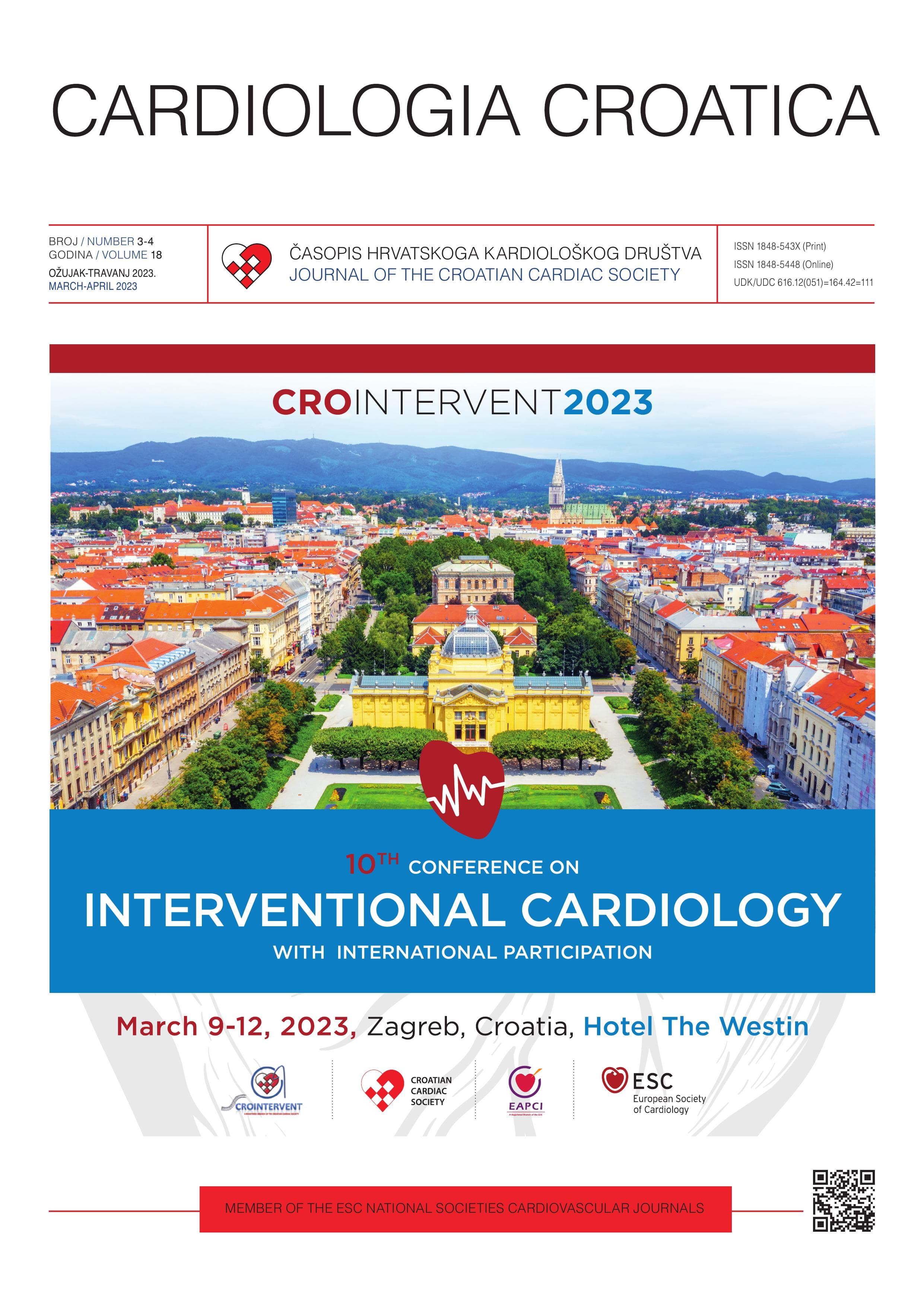
## Dear Colleagues, It is a true pleasure and honor to invite you to the 10th Croatian Conference on Interventional Cardiology – CROINTERVENT 2023, which will be held in Zagreb, on March 9-12, 2023. Almost two years have passed since the last congress, which was the first cardiology congress that was fully held in person after the easing of the epidemiological situation related to the COVID-19 pandemic. The meeting confirmed the incalculable value of direct communication during the exchange of knowledge and experiences. We will retain the well-demonstrated and established concept of a congress that provides an overview of the current achievements in diagnostic and therapeutic procedures applied in modern interventional cardiology. The congress program will include invited lectures by eminent domestic and foreign experts, as well as oral and moderated poster presentations. In addition to theoretical knowledge, as always, an important part of the congress will be devoted to practical aspects of interventional cardiology. Direct video transmissions of procedures from interventional laboratories will remain a recognizable and very important part of the congress, and cases of previously performed procedures will also be presented with discussion and comments. A significant part of the congress will be devoted to pharmacotherapy as a vital part of interventional cardiology, which has proven its value both in primary and secondary prevention of cardiovascular diseases, as well as in improving primary outcomes and reducing the frequency of complications of cardiovascular interventions. Faithful to its core concept, CROINTERVENT 2023 remains aimed not only at cardiologists but also towards physicians of other specialties, as well as all others who are professionally associated with this important segment of modern cardiology. The goal of the congress is to present the current state and possibilities of modern interventional cardiology, identify areas that are still under-researched, harmonize positions on controversial topics, consider optimal treatment strategies in the future, and to discuss the difficulties that we face in clinical practice every day. This Supplement of Cardiologia Croatica – the official journal of the Croatian Cardiac Society – consists of selected original contributions from our participants in the form of abstracts, which will be presented at the meeting in the form of oral presentations and moderated posters. Sincerely yours, Doc. dr. sc. Eduard Margetić / Assist Prof Eduard Margetić, MD, PhD Predsjednik Radne skupine za invazivnu i intervencijsku kardiologiju Hrvatskog kardiološkog društva / President, Working Group on Invasive and Interventional Cardiology of the Croatian Cardiac Society
Nikolina Glavinić, Bruno Mihatović, Ana Dadić, Mihael Matić
**Introduction**: Antiphospholipid syndrome is an autoimmune coagulation disorder that is manifests clinically as recurrent venous or arterial thrombosis. In some case thrombocytopenia can be the only manifestation of the syndrome. (1-3) **Case report**: We present a 43-year-old patient with previously known antiphospholipid syndrome who was admitted to the Coronary Care Unit due to acute myocardial infarction with ST-elevation of the posterolateral location (STEMI). An emergency coronary angiography was performed, which showed thrombotic occlusion of the proximal segment of the circumflex artery. A primary percutaneous coronary intervention was performed, and the target lesion was treated with drug coated balloon (DCB)-Sequent Please Neo 3.0x20 mm. In the control angiography, there is no narrowing. There is a normal TIMI grade 3 flow without any residual thrombus. In the literature, results after treatment of ST-elevation acute coronary syndrome in patients with antiphospholipid syndrome are variable. When PCI is used for STEMI in patient with antiphospholipid syndrome, DCB therapy (although not mentioned in current guidelines) is safe and effective and has shown good clinical effects during a one-year follow-up period. Dual antiplatelet therapy (clopidogrel + aspirin) was started along with warfarin with a target INR >2 (The indication for anticoagulant therapy is the previously known antiphospholipid syndrome). After achieving target INR values, he was discharged and referred to rheumatology and cardiology outpatient clinics for further follow-up. For patients experiencing an acute myocardial infarction of thrombophilic genesis, using thromboaspiration, antiplatelet and anticoagulant medications, as well as applying drug- coated balloons (DCB), can help avoid the placement of a stent and all the complications associated with it. The BASKET SMALL 2 trial has described how this method can provide optimal results.
Katica Cvitkušić Lukenda, Marijana Knežević Praveček, Ivica Dunđer, Antonija Raguž, Ivan Bitunjac, Domagoj Mišković, Jelena Jakab, Ivana Grgić, Blaženka Miškić, Branka Bardak, Anto Lukenda, Krešimir Gabaldo
**Introduction**: Primary percutaneous coronary intervention (PCI) is recommended for patients with ST-segment elevation myocardial infarction (STEMI) if it can be performed in a timely manner. Universal access is the major limitation of a PCI strategy, especially for STEMI patients who are usually initially transferred to a non-PCI hospital. A PCI network-oriented approach that bypasses non PCI centers in favor of PCI capable centers can reduce system delay and improve patient outcomes. (1-3) **Patients and Methods**: The General Hospital “Dr Josip Benčević” is a PCI-center in Western Slavonia within the Croatian PCI Network. We have developed a regional protocol for transfer of field-triaged patients directly to our hospital, bypassing local non-PCI hospitals. The Cath Lab was notified by the field emergency medical service (EMS) when a STEMI was suspected. Premedication with a loading dose of aspirin and a P2Y12 inhibitor was administered according to established protocol, and patients were admitted directly to the Cath Lab. Patient- and procedure-specific information as well as data from the Hospital Information Service and EMS register were documented in our register. **Results**: Between January 2020 and October 2021, a total of 37 patients with suspected STEMI infarction were transferred to the Cath Lab, including 13 women with a mean age of 75 years (53-91 years) and 24 men with a mean age of 61 years (46-80 years). STEMI diagnosis was confirmed in 33 patients, 2 patients had pericarditis, 1 patient had Takotsubo cardiomyopathy, and 1 patient had hypertensive crisis. The culprit artery was he right coronary artery (RCA) in 17 (51.5%) patients, left anterior descending artery in 9 (27.3%) patients, circumflex artery in 5 (15.2%) patients, venous bypass graft in 1 (3%) patient, and 1 (3%) patient refused the procedure. All patients received aspirin, 14 (42.4%) patients received a P2Y12 inhibitor. Two patients suffered cardiac arrest during transport due to malignant arrhythmias (VF/VT). Patients with RCA occlusion were transported faster (median 37 minutes) than those with LAD occlusion (median 41 minutes). **Conclusion**: Direct communication between EMS and field-triaging patients to the PCI center resulted in safe and efficient transport of patients to the cath lab. A shorter health care system delay can reduce reperfusion time and may be the key to further improving cardiovascular outcomes in STEMI patients.