Cardiologia Croatica
Journal Research Assistant
Journal Research Assistant
Journal Research Assistant
Journal Research Assistant
Sergej Nadalin, Domagoj Vučić, Marko Galić, Josip Silović, Jadranka Vraneković, Ivan Majdandžić, Darko Margetić, Katica Cvitkušić Lukenda
Folate is an essential form of vitamin B9 that facilitates the transfer of single-carbon groups and plays an important role in DNA methylation, conversion of homocysteine to methionine, nucleotide synthesis, and DNA repair. Folate deficiency has been reported to be associated with an increased risk of coronary artery disease (CAD). Although the biochemical mechanisms linking folate deficiency to CAD have been extensively studied, the genetics of folate metabolism and the clinical implications of folate supplementation remain areas of ongoing investigation. We integrated three interconnected domains—biochemistry, genetics, and nutritional supplementation—to provide a comprehensive synthesis of current knowledge on the folate metabolism in CAD. There is strong evidence that elevated plasma homocysteine levels are associated with increased cardiovascular mortality and long-term adverse events in patients with CAD, often due to folate deficiency. Elevated plasma homocysteine levels contribute to endothelial dysfunction, oxidative stress, inflammation, and atherosclerotic plaque formation, all of which play roles in pathogenesis of CAD (1). The genes encoding the enzymes methylenetetrahydrofolate reductase (MTHFR) and methyltetrahydrofolate-homocysteine methyltransferase reductase (MTRR) play a central role in the metabolism of folate, homocysteine, and consequently in methylation. The MTHFR gene is involved in the conversion of methylenetetrahydrofolate to methyltetrahydrofolate, which is the main circulating form of folate and the carbon donor for the remethylation of homocysteine to methionine. Meanwhile, MTRR gene plays a role in the remethylation of homocysteine to methionine via cobalamin in a folate-dependent reaction. Polymorphisms in the MTHFR and MTRR genes can influence the risk of CAD by altering the metabolism of homocysteine. Of particular importance is the MTHFR C677T polymorphism, while the A1298C polymorphism and, especially, the MTRR A66G polymorphism have shown less consistent associations with CAD risk (2, 3). In addition, folate supplementation lowers homocysteine levels, particularly in individuals with the MTHFR C677T TT genotype, which is associated with reduced enzyme activity involved in homocysteine metabolism (4). Importantly, although folic acid supplementation reduces plasma homocysteine, systematic reviews and meta-analyses show no significant effect on CAD risk or cardiovascular mortality. These findings highlight the need for further research to better understand the mechanisms and conditions under which folate supplementation might offer cardiovascular protection.
Nataša Moser
The importance of proper nutrition, encompassing both dietary diversity and a balanced intake of nutrients, is now indisputable. In the modern food pyramid, the position of fats has undergone a remarkable transformation compared to that of the 1990s, shifting from the top to the bottom. This change reflects a growing body of evidence emphasizing the fundamental significance of both the quantity and quality of dietary fats (1, 2). Those fats form the lipid pool that serves as a source of fats for many essential physiological functions, for example, they are fundamental components of cellular membranes, forming the hydrophobic tails of phospholipid bilayers. The completer and more balanced the lipid pool, the better the structural and functional integrity of the cell membrane. The quality and proportion of lipids within membranes determine not only membrane fluidity and permeability but also the efficiency of cellular metabolism. Just as maintaining a balanced dietary intake of saturated, monounsaturated, and polyunsaturated fatty acids is essential, their proportional balance within the membrane structure is equally critical. An excessive amount of either saturated or unsaturated fatty acids disrupts the integrity of the cell membrane due to its rigidity and permeability and stimulates cholesterol synthesis as a compensatory mechanism. The relative proportion of polyunsaturated fatty acids, particularly omega-6 and omega-3, also influences inflammatory potential. During membrane remodeling, these fatty acids are released and metabolized into pro- or anti-inflammatory mediators. While inflammation is an essential defensive mechanism involved in immune responses and tissue repair, an excess of pro-inflammatory mediators, because of an excess of their precursors, such as arachidonic acid (omega-6), may lead to a self-sustaining inflammatory cycle. This uncontrolled process can result in chronic inflammation, a key contributor to many metabolic and degenerative diseases (1, 3). Cell membranes and their fatty acid composition are therefore crucial determinants of overall cellular metabolism. The molecular characteristics of the membrane should not be neglected in clinical practice, as its lipid profile reflects both intrinsic biosynthetic capacity and long-term dietary habits.
Adrijana Inđić, Dragan Karan, Lejla Bešić, Slađana Marčeta
In the cardiac catheterization laboratory, precis and controlled administration of contrast media plays a crucial role in ensuring high diagnostic image quality while minimizing the risk of adverse effect in patients. Traditionally contrast has been injected manually using a syringe, which may lead to variation in the amount and rate of injection depending on the operator’s experience and technique. Modern automatic injectors represent a technological advancement that enables accurate dosing, constant over the quantity of contrast administered. (1) The aim of this study is to compare the consumption of contrast media during cardiac catheterization procedures performed with manual injection versus those performed using an automatic injection. The analysis will be based on data collected from the hospital information system, including procedure type, procedure duration, injection protocol, and patient characteristics. Special attention will be given to the total amount of contrast used per procedure and the potential for dose optimization. It is expected that the results will demonstrate a reduction in overall contrast consumption when using an automatic injector, while maintaining diagnostic image quality. Such an outcome may have significant clinical and economic implications, as reduced contrast use could contribute to lowering the risk of contrast-induced nephropathy, shortening procedure duration, and promoting more rational use of materials in catheterization laboratories. The findings of this study could serve as a basis for standardizing the use of automatic injectors in interventional cardiology, with the potential to improve patient safety and the efficiency of healthcare staff.
Zvonimir Bosnić, Matija Vuksanović, Iva Petričušić, Mislav Omerbašić, Domagoj Vučić, Katica Cvitkušić-Lukenda
The immunometabolic interface represents a crucial link between metabolic dysfunction and chronic inflammation. Chronic low-grade inflammation is increasingly recognized as a key driver of metabolic, cardiovascular, and renal disorders. In the pathophysiology of diabetes mellitus and its complications—including cardiovascular disease and nephropathy—activation of the nuclear factor kappa B (NF-κB) signaling pathway plays a central role. Triggered by pro-inflammatory cytokines and damage-associated molecular patterns, this pathway promotes sustained inflammatory responses, β-cell dysfunction, and the development of insulin resistance. Furthermore, activation of the NLRP3 inflammasome, mediated by mitochondrial dysfunction and impaired autophagy, amplifies the inflammatory cascade through the release of interleukin (IL)-1 and IL-18. These processes together establish a chronic pro-inflammatory state that contributes to metabolic deterioration and vascular injury (1, 2). Recent advances in antidiabetic therapy have highlighted that several novel drug classes, initially developed for glycemic control, possess significant immunomodulatory and anti-inflammatory properties. Sodium-glucose cotransporter-2 (SGLT2) inhibitors improve vascular health by attenuating oxidative stress and inflammation, enhancing endothelial function, modulating microRNA expression, and delaying vascular aging and atherosclerosis (1). Dipeptidyl peptidase-4 (DPP-4) inhibitors exert anti-inflammatory effects by suppressing NF-κB activation, reducing pro-inflammatory cytokines, and limiting macrophage and T-cell infiltration, thereby improving endothelial function and stabilizing atherosclerotic plaques. Similarly, glucagon-like peptide-1 (GLP-1) receptor agonists reduce systemic inflammation through downregulation of CRP, TNF-α, IL-1, and IL-6, while also inhibiting NF-κB signaling. By mitigating vascular inflammation and oxidative stress, these agents improve cardiovascular outcomes and reduce heart failure–related hospitalizations (2). These findings highlight the therapeutic significance of targeting immunometabolic and inflammatory pathways to preserve vascular and renal integrity. By attenuating chronic low-grade inflammation and oxidative stress, such interventions may not only prevent cardiovascular events and slow chronic kidney disease progression but also provide a framework for developing future therapies targeting inflammation-driven complications in type 2 diabetes.
Karlo Petošić, Marijana Knežević Praveček
The use of telemedicine in prehospital care of STEMI patients in Brodsko-Posavska County enables timely recognition and activation of the intervention team in the percutaneous coronary intervention center, which directly contributes to a shorter time from first medical contact to reperfusion. The high rate of aspirin administration, timely venous access and 12-lead electrocardiogram within the recommended time frame indicate good adherence to clinical guidelines in the daily work of emergency medical teams. However, differences in the availability of teams with medical staff and logistical challenges in remote areas remain obstacles to achieving optimal time intervals. The integration of highly educated nurses and technicians with specialized competencies in emergency medicine represents a significant advance and offers the potential for further improvements in the quality of care, especially in teams without the constant presence of a physician. (1-3) Further research is needed to monitor treatment outcomes and assess the effectiveness of current protocols.
Marija Divić, Katarina Ćurić, Ivica Dunđer, Krešimir Gabaldo, Domagoj Mišković, Katica Cvitkušić Lukenda, Marijana Knežević Praveček
In order to improve the availability of specialist cardiology care at the primary healthcare level, a cardiology outpatient clinic was established in 2022 within the Health Centre Brod-Posavina County. The clinic operates in cooperation with five cardiologists from the General Hospital “Dr Josip Benčević”, enabling early detection, diagnosis, and monitoring of cardiac patients without the need for hospital referrals. (1, 2) The nurse plays a key role in the daily organization of the clinic. Responsibilities include scheduling patients for consultations and diagnostic procedures such as 12-lead electrocardiogram, blood pressure measurement, 24-hour Holter ECG and blood pressure monitoring, exercise stress testing, and cardiac ultrasound. The nurse also organizes follow-up appointments, maintains patient records, and ensures timely communication regarding appointments and preparation for procedures. In the period from September 2022 to September 2025, a total of 1.039 patients were examined. The following procedures were performed: 1.376 24-hour ECG Holter monitorings, 451 blood pressure Holter monitorings, 968 echocardiograms, 237 exercise stress tests and 1.051 standard ECGs. One of the nurse’s most important tasks is ensuring effective communication and collaboration between family medicine teams and cardiology specialists. This facilitates timely diagnosis and treatment and reduces the need for hospital admissions. The role of the nurse in this model goes beyond traditional nursing duties and includes workflow coordination, communication with patients and healthcare professionals, as well as patient education regarding medication adherence and lifestyle changes. Experience from the first three years of clinic operation indicates that this organizational model significantly improves access to and the quality of cardiology care at the primary level, with high patient satisfaction.
Darko Adžić
The use of vascular closure devices (VCDs) has become an essential component in modern interventional procedures, offering a faster, safer, and more efficient method for achieving hemostasis following catheterization. This study/presentation focuses on the design, mechanism, and application of closure devices, emphasizing their role in improving patient outcomes and workflow efficiency for medical technicians. The device functions by mechanically sealing the arterial puncture site using bioabsorbable materials or suture-based systems, thereby reducing manual compression time, minimizing patient discomfort, and enabling earlier ambulation. Key procedural considerations, including device selection, deployment technique, and post-procedural monitoring, are discussed to enhance clinical proficiency and safety. Understanding the principles of closure device operation allows medical technicians to assist effectively during vascular interventions and to identify and manage potential complications such as bleeding, infection, or device failure. Mastery of these techniques ultimately contributes to optimal patient care and procedural success in cardiovascular and endovascular settings. (1, 2)
Ivana Vučinić Ljubičić, Hrvoje Holik, Božena Coha, Stjepan Kovačević
**Introduction:** Bruton’s tyrosine kinase inhibitors (BTKi) have revolutionized the management of chronic lymphocytic leukemia (CLL), offering effective chemotherapy-free options across different treatment settings (1). However, first-generation BTKi ibrutinib is associated with increased cardiovascular toxicity, particularly atrial fibrillation and hypertension (1-3). Second-generation BTKi acalabrutinib and zanubrutinib were developed to enhance selectivity and reduce occurrence of these events, which has been confirmed in both clinical trials and recent real-world analyses (2-4). Our aim was to compare the incidence of treatment-emergent arterial hypertension and atrial fibrillation among patients with CLL treated with first- versus second-generation BTKi. **Patients and Methods**: We retrospectively analyzed 30 adult CLL patients treated with BTKi in our Hematology Department between December 2017 and December 2024. Seventeen patients received ibrutinib and thirteen patients a second-generation BTKi (acalabrutinib or zanubrutinib). Clinical data, including demographics and cardiovascular comorbidities were collected from electronic medical records. Hypertension and atrial fibrillation were defined and graded according to CTCAE v5.0. Baseline hypertension was defined as a previously known condition or ongoing therapy; de novo cases and worsening events were identified during treatment. **Results:** At treatment initiation, arterial hypertension was present in 59% of patients receiving ibrutinib and in 62% of those treated with second-generation BTKi (**Table 1**). During therapy, de novo hypertension occurred in 18% of ibrutinib-treated patients compared with 8% among those receiving second-generation BTKi, while worsening of pre-existing hypertension was observed in 6% and 8% of patients, respectively. None of the patients had a history of atrial fibrillation at baseline. During treatment, de novo atrial fibrillation developed in 3 patients (18%) on ibrutinib and in 1 patient (8%) receiving second-generation BTKi. Most cardiovascular events were grade 1–2 and managed with standard therapy without permanent discontinuation of BTKi treatment. ### TABLE 1: Baseline characteristics and treatment-emergent cardiovascular events in patients with chronic lymphocytic leukemia treated with first- and second-generation Bruton’s tyrosine kinase inhibitors. | **Baseline characteristics** | **All patients (N=30)** | **First-generation BTKi (ibrutinib) (n=17)** | **Second-generation BTKi (acalabrutinib / zanubrutinib) (n=13)** | | --- | --- | --- | --- | | **Demographics** | | | | | Gender M/F | 16 (53%) / 14 (47%) | 10 (59%) / 7 (41%) | 6 (46%) / 7 (54%) | | Age (median/range) | 73 / 56-88 y | 72 / 56-83 y | 75 / 59-88 y | | **Clinical characteristics** | | | | | Hypertension at baseline, n (%) | 18 (60%) | 10 (59%) | 8 (62%) | | -De novo hypertension during therapy, n (%) | 4 (13%) | 3 (18%) | 1 (8%) | | - Worsening hypertension, n (%) | 2 (7%) | 1 (6%) | 1 (8%) | | Atrial fibrillation at baseline, n (%) | 0 (0%) | 0 (0%) | 0 (0%) | | - De novo atrial fibrillation, n (%) | 4 (13%) | 3 (18%) | 1 (8%) | [†] Values are presented as number (percentage) or median (range), as appropriate. Baseline hypertension and baseline atrial fibrillation denote previously known diagnoses or ongoing therapy at BTKi initiation. De novo hypertension and de novo atrial fibrillation refer to new-onset events occurring during BTKi therapy in patients without these conditions at baseline. Worsening hypertension was defined as an increase of ≥ 1 CTCAE v5.0 grade or the need for intensification of antihypertensive therapy. Abbreviations: BTKi – Bruton’s tyrosine kinase inhibitor; AF – atrial fibrillation; CTCAE – Common Terminology Criteria for Adverse Events. **Conclusion:** In this single-centre real-world analysis, cardiovascular adverse events, particularly hypertension and atrial fibrillation, were more frequent in patients treated with ibrutinib compared to those receiving second-generation BTKi. These findings support previous evidence suggesting a more favorable cardiovascular safety profile of acalabrutinib and zanubrutinib, emphasizing the importance of systematic cardiovascular monitoring and multidisciplinary collaboration between hematologists and cardiologists during BTKi therapy.
Zvonimir Bosnić, Matija Vuksanović, Blaženka Šarić, Tatjana Bačun, Iva Petričušić, Mislav Omerbašić
Age-related metabolic and vascular diseases—such as type 2 diabetes, obesity, hypertension, and atherosclerosis—are leading contributors to morbidity and mortality worldwide. While traditionally studied in isolation, these conditions share overlapping pathophysiological mechanisms, particularly at the intersection of immune and metabolic dysfunction. Increasing evidence supports the existence of common immune-metabolic disturbances, including chronic low-grade inflammation (inflammaging), immune cell dysregulation, mitochondrial dysfunction, and altered nutrient sensing (1). These factors contribute to endothelial dysfunction, insulin resistance, oxidative stress, and tissue remodeling—the hallmarks of both metabolic and vascular decline in aging. Despite significant advances in immunometabolism and aging biology, our understanding of how these immune-metabolic alterations develop and interact during aging remains incomplete. Key shared pathways—such as activation of the NLRP3 inflammasome, impaired AMPK and mTOR signaling, macrophage polarization, and immune senescence—have been identified (2). However, the causal relationships between these processes and their temporal dynamics across different tissues are still poorly defined. Moreover, heterogeneity in aging phenotypes, sex differences, and the influence of environmental and genetic factors further complicate the elucidation of unifying mechanisms. This lack of integrative understanding presents a major barrier to the development of effective, targeted interventions for age-related cardiometabolic diseases (3). Current therapeutic strategies often address individual risk factors rather than the underlying systemic dysfunctions. There is an urgent need for interdisciplinary approaches that combine systems biology, longitudinal studies, and mechanistic research to map the complex crosstalk between the immune and metabolic systems during aging. In this review, we synthesize current knowledge on common immune-metabolic mechanisms driving age-related metabolic and vascular conditions, highlight key gaps in the field, and propose future research directions. A deeper understanding of these shared pathways could pave the way for innovative therapies aimed at delaying or preventing multiple age-associated diseases through immune-metabolic modulation.
Mario Špoljarić, Katica Cvitkušić Lukenda, Kristina Vorkapić, Marko Galić, Matea Kirchbauer Špoljarić, Dijana Bešić, Anđela Jurišić
**Introduction**: Systemic infections, particularly acute respiratory infections, are recognized triggers of acute coronary syndromes (ACS). They may precipitate plaque destabilization through systemic inflammation, endothelial dysfunction, and platelet activation, thereby lowering the threshold for plaque rupture or erosion. (1, 2) **Case report:** 59-year-old woman with no prior comorbidities was admitted to the Coronary Care Unit with acute heart failure following five days of persistent chest pain. During the same period, she experienced sustained fever accompanied by chills, rigors, and a dry cough. On admission, the patient was dyspneic and orthopneic and reported mild pressure in the left hemithorax. Laboratory tests showed elevated high-sensitivity troponin I (3546 ng/L), N-terminal pro-B-type natriuretic peptide (6225 pg/mL), and inflammatory markers (C-reactive protein 125 mg/L, leukocytes 11×109/L). Serial measurements demonstrated a gradual decline in hsTnI levels. Electrocardiography revealed ST-segment elevation in leads V1–V4 with Q-wave formation, while echocardiography confirmed akinesia of the apical, interventricular septal, and anterior walls, reduced left ventricular ejection fraction, and an apical thrombus (**Figure 1**). Given the concurrent fever and chest pain, acute myocarditis was initially included in the differential diagnosis. Chest X-ray showed extensive bilateral pneumonia, confirmed by MSCT pulmonary angiography (**Figure 2**). Coronary angiography performed the following day revealed occlusion of the ostial left anterior descending artery with heterocollateralization from other epicardial vessels, consistent with a subacute anterior ST-elevation myocardial infarction (**Figure 3**). The patient received broad-spectrum intravenous antibiotics and optimal pharmacological therapy for heart failure. Gradual cardiac recompensation was observed, with resolution of chest pain and a marked decline in inflammatory markers. She was discharged on the seventh day of hospitalization on optimized pharmacotherapy for coronary artery disease and heart failure, including anticoagulation with warfarin. Myocardial viability assessment is planned three months post-discharge to guide further interventional management. FIGURE 1. Contrast-enhanced echocardiogram demonstrates a left ventricular apical thrombus. FIGURE 2. The scan shows bilateral, predominantly peripheral ground-glass opacities with patchy consolidation, consistent with pneumonia. FIGURE 3. Ostial left anterior descending artery (LAD) occlusion with collateral flow from the circumflex artery. **Conclusion**: This case highlights how acute infection and systemic inflammation can mask or mimic acute coronary syndromes, complicating the diagnosis and delaying definitive treatment. Early coronary evaluation should be considered in febrile patients presenting with chest pain and elevated cardiac biomarkers to ensure timely management and improve clinical outcomes.
Josip Silović, Domagoj Mišković, Ivan Majdandžić, Marijana Knežević Praveček, Katica Cvitkušić Lukenda
**Introduction**: Modern cardiac catheterization laboratories and coronary care units (CCU) require sophisticated sedation protocols that balance patient comfort with hemodynamic stability and respiratory safety (1). This review presents sedation options currently implemented at General Hospital Slavonski Brod’s CCU. **Protocols and Methods**: We analyzed seven primary sedation agents used in our institution: propofol and propofol TCI (Target-Controlled Infusion), remifentanil TCI, dexmedetomidine, sufentanil, fentanyl, and midazolam. Each agent’s pharmacokinetics, pharmacodynamics, indications, contraindications, and adverse effects were systematically reviewed based on current literature and institutional experience (**Table 1** and **Table 2****).** Our sedation protocols are tailored to specific procedures: propofol for electrical cardioversion due to rapid onset and recovery (2); midazolam combined with propofol for transcatheter aortic valve implantation procedures; dexmedetomidine for mechanically ventilated patients post-cardiac arrest or myocardial infarction; and dexmedetomidine-based protocols for anxious elderly patients and non-invasive ventilation (NIV) mask tolerance enhancement (3, 4). Each approach considers patient-specific factors including age, comorbidities, and procedural complexity. Propofol demonstrates rapid onset (30-40 seconds) with significant cardiovascular depression but predictable recovery. TCI systems provide superior concentration control and reduced side effects. Remifentanil TCI offers precise analgesia control with ultra-short elimination half-life (3-10 minutes) but requires vigilant respiratory monitoring. Dexmedetomidine provides unique conscious sedation with minimal respiratory depression, making it ideal for prolonged sedation and NIV tolerance (3, 4). Traditional opioids (fentanyl, sufentanil) and midazolam remain valuable for specific indications with established safety profiles. Each agent presents distinct contraindication patterns. Propofol requires caution in cardiac failure and hypovolemia (2). Dexmedetomidine necessitates monitoring for bradycardia and hypotension. Opioids demand respiratory surveillance, while midazolam may cause paradoxical reactions in elderly patients. ### TABLE 1: Contraindications and special warnings. | **Drug** | **Absolute contraindications** | **Relative contraindications** | **Special warnings** | | --- | --- | --- | --- | | Propofol | Allergy to propofol, eggs, soy | Heart failure, hypovolemia | Propofol infusion syndrome, hypertriglyceridemia | | Remifentanil TCI | Allergy to fentanyl analogues | Severe renal/hepatic insufficiency, COPD, mechanical obstruction of the GIT | Muscle rigidity, respiratory depression | | Dexmedetomidine | No absolute contraindications | Bradycardia <50/min, hypotension, heart block | Bradycardia, prolonged action in the elderly | | Sufentanil/ Fentanyl | Allergy to opioid analgesics, acute asthma | CNS depression, increased intracranial pressure | Respiratory depression, addiction | | Midazolam | Allergy to benzodiazepines, acute glaucoma | Dementia, COPD, myasthenia gravis | Delirium in the elderly, anterograde amnesia | [†] TCI – Target-Controlled Infusion; COPD – Chronic Obstructive Pulmonary Disease; CNS – Central Nervous System; GIT – Gastrointestinal Tract ### TABLE 2: Pharmacokinetics and pharmacodynamics. | Drug | **Mechanism of action** | **Onset of action** | **Elimination half-time** | **Cardiovascular effects** | **Respiratory effects** | | --- | --- | --- | --- | --- | --- | | Propofol | GABA receptor agonist | 30-40 seconds | 4-7 hours | Hypotension, ↓CO, bradycardia | Respiratory depression, apnea | | Propofol TCI | GABA receptor agonist | 30-40 seconds | 4-7 hours | Hypotension, ↓CO, bradycardia | Respiratory depression, apnea | | Remifentanil TCI | μ-opioid receptor agonist | 1-3 minutes | 3-10 minutes | Bradycardia, hypotension | Significant respiratory depression | | Dexmedetomidine | α2-adrenoreceptor agonist | 15 minutes (without loading dose) | 2-3 hours | Bradycardia, hypotension, initial hypertension | Minimal respiratory depression | | Sufentanil | μ-opioid receptor agonist | 1-3 minutes | 2.5-3 hours | Bradycardia, mild hypotension | Significant respiratory depression | | Fentanyl | μ-opioid receptor agonist | 1-2 minutes | 3-4 hours | Bradycardia, mild hypotension | Moderate respiratory depression | | Midazolam | GABA-A receptor agonist | 1-3 minutes | 1-4 hours | Minimal | Mild respiratory depression | [†] TCI – Target-Controlled Infusion; ↓CO – Decreased Cardiac Output; GABA – gamma-aminobutyric acid **Conclusions**: Successful sedation in cardiac catheterization laboratories and coronary units require individualized approaches based on pharmacological understanding, procedural requirements, and patient characteristics. Our institutional experience demonstrates that combined protocols utilizing multiple agents can optimize patient outcomes while maintaining safety. Continuous monitoring and staff education remain paramount for safe sedation practice.
Matilda Pudić, Ivica Dunđer, Josip Silović
**Introduction**: Antiphospholipid syndrome (APS) is a rare autoimmune disorder characterized by increased propensity to thrombotic events, pregnancy complications and persistent presence of antiphospholipid antibodies (1). Although relatively uncommon, APS has significant clinical importance due to potentially life-threatening complications. In APS patients, thrombotic events represent the most common syndrome manifestation. Deep vein thrombosis with pulmonary embolism is the most common manifestation (2). APS can be primary (isolated) or secondary, associated with other autoimmune diseases, most commonly systemic lupus erythematosus (SLE), where 30-40% of SLE patients have antiphospholipid antibodies (3). **Case report**: 22-year-old male patient, who presented to the Emergency Department with dyspnea during physical activity, followed by collapse and loss of consciousness. MSCT pulmonary angiography confirmed thromboembolic disease of large pulmonary artery branches bilaterally, with pericardial effusion. Treatment was immediately initiated with therapeutic-dose low-molecular-weight heparin. From the sixth day of hospitalization, peroral anticoagulant therapy with warfarin was introduced. Laboratory findings showed a positive ANA titer 1:320 with cytoplasmic and punctate pattern, elevated anti-dsDNA (31 H), and positive beta-2 glycoprotein I antibodies (37.9). Lupus anticoagulant was negative. These findings combined with clinical presentation confirmed antiphospholipid syndrome diagnosis. Multidisciplinary treatment approach included rheumatological follow-up with introduced hydroxychloroquine 200 mg daily, along with regular hematologist and cardiologist controls. Continuation of anticoagulant therapy and serological marker monitoring is recommended. Follow-up immunological workup is planned three months after initial testing to confirm antiphospholipid antibody persistence. **Conclusion**: This case illustrates APS as a significant diagnosis in young adults with unexplained pulmonary embolism. Early diagnosis and timely anticoagulant treatment are crucial for preventing recurrent thrombotic events. A multidisciplinary approach with regular monitoring enables optimal management of this complex autoimmune disorder.
Ines Novak, Marina Baličević, Ivica Čabraja
**Introduction**: Infective endocarditis (IE) in a patient with Marfan syndrome and a mechanical mitral valve prosthesis presents both diagnostic and therapeutic challenges, particularly in cases of culture-negative endocarditis. Timely recognition and empiric antimicrobial therapy are essential for achieving a favorable clinical outcome. (1-3) **Case report:** 52-year-old male with a known history of Marfan syndrome, previous mechanical mitral valve replacement, Valsalva aortic graft implantation and radiofrequency ablation of arrhythmogenic foci in the left atrium, was admitted due to a one-month history of low-grade fever, rhinorrhea and worsening dyspnea. The onset of symptoms occurred six months postoperatively. Clinical examination, laboratory tests, serological analyses and imaging studies failed to identify focus of infection and all obtained blood culture sets (including HACEK organisms) remained sterile. Transthoracic echocardiography (TTE) did not reveal any vegetations or intracavitary masses; however, transesophageal echocardiography (TEE) demonstrated a hyperechogenic lesion measuring 0.4 × 0.4 cm on the non-coronary cusp of the aortic valve. Positron emission tomography–computed tomography (PET-CT) revealed increased radiotracer uptake in the ascending aorta, accompanied by mild morphological irregularity of the valve, consistent with an active inflammatory process. Based on the 2023 Duke criteria, a diagnosis of definitive infective endocarditis was established. Empirical combination therapy with meropenem, vancomycin and rifampicin were initiated and continued for 6 weeks, after which the patient became afebrile and clinically stable. He was subsequently referred for outpatient cardiology follow-up. **Conclusion**: Despite advances in diagnostic modalities, culture-negative endocarditis remains a significant clinical challenge due to the difficulty in differentiating infectious from sterile inflammatory lesions. In this context, PET-CT has emerged as a valuable adjunctive imaging technique, aiding in the confirmation of infection in diagnostically ambiguous cases. This case highlights the complexity of diagnosing culture-negative endocarditis in a patient with Marfan syndrome and multiple prior cardiovascular surgeries. In the absence of microbiological confirmation, multimodal imaging (TTE, TEE, PET-CT) combined with timely empiric antimicrobial therapy remains crucial for successful management and prevention of potential complications associated with infective endocarditis.
Antonia Majetić, Marijana Knežević Praveček, Barica Stanić
**Introduction:** Health literacy is a critical factor in the management of cardiovascular diseases and has a direct impacts patients’ quality of life. Understanding the relationship between health literacy and quality of life can help identify patient groups at risk and guide targeted interventions. (1-4) This study aimed to assess health literacy and quality of life in patients with cardiovascular diseases, and to explore their association, while considering differences in gender, age, education, marital status, and place of residence. **Patients and Methods:** A cross-sectional study was conducted at the Department of Cardiology, General Hospital “Dr. Josip Benčević,” between March and May 2025. Hospitalized patients with cardiovascular diseases were included. Data were collected using an anonymous written questionnaire including sociodemographic questions, the SAHLSA-50 test for health literacy, and the SF-36 questionnaire for quality of life. Due to non-normal data distribution, nonparametric tests (Mann–Whitney, Kruskal–Wallis with post hoc analysis) were applied. Significance was set at P < 0.05. Analyses were performed using JASP version 0.19.3. **Results:** Participants showed moderate health literacy, with no significant differences across most demographic groups. Mental health scored highest, whereas physical functioning exhibited the greatest limitations. Higher health literacy was correlated with better overall quality of life and fewer emotional limitations. Significant differences in literacy and quality of life were observed with respect to age, education, and place of residence. Men reported better physical functioning, while women experienced greater emotional limitations. Older participants reported more intense pain. **Conclusion:** Health literacy among patients with cardiovascular diseases is generally moderate and positively influences quality of life, particularly in mental health and physical functioning. Demographic factors including age, education, gender, and place of residence significantly affect both health literacy and quality of life. These findings highlight the importance of tailored interventions to improve patient outcomes in cardiovascular care.
Ana Kovačević, Stjepan Kovačević, Iva Dumančić, Marijana Kovačević, Maja Franić, Josipa Meter, Nikolina Bukal, Ninoslav Leko, Katica Cvitkušić Lukenda
**Introduction:** Smoking is a well-established risk factor for the development of acute coronary syndrome (ACS). (1) Beyond its role in disease onset, smoking has been associated with adverse outcomes during follow-up. (2) While several studies have examined the impact of smoking cessation on cardiac recovery, few have directly compared smokers and non-smokers with respect to functional cardiac recovery following ACS. (3) The aim of this study was to evaluate differences in cardiac functional recovery between smokers and non-smokers after ACS. **Patients and Methods:** This retrospective study included patients hospitalized with ACS between January and December 2023. Patients were classified as smokers or non-smokers. Variables included demographics, comorbidities, MACE score, troponin, stenosis extent, hypokinesia, diastolic dysfunction, and echocardiographic changes. Analyses were performed using SPSS 26. Categorical variables were compared with Fisher’s exact or Chi-square tests, and continuous variables with Student’s t-test or Mann–Whitney U test. A two-tailed p 0.99† | | Hyperlipidemia | 17 (53.1) | 15 (46.9) | 32 (76.2) | 0.5 | | Hypertension | 13 (46.4) | 15 (53.6) | 28 (66.7) | 0.51 | | Diabetes mellitus | 5 (83.3) | 1 (16.7) | 6 (14.3) | 0.18† | | COPD | 2 (100) | 0 (0) | 2 (4.8) | 0.49† | [†] *χ2 test; †Fisher’s exact test ### TABLE 2: Comparison of cardiac recovery between smokers and non-smokers after acute coronary syndrome. | | **Median (IQR)** — **Smokers** | **Median (IQR)** — **Non-smokers** | **P*** | | --- | --- | --- | --- | | LVEF (%) | | | | | Baseline value | 55 (45-62) | 57 (45-60) | 0.6 | | Follow-up value | 60 (52-62) | 60 (49-67) | 0.6 | | Left ventricle (cm) | | | | | Baseline value | 51 (45-53) | 52 (46-56) | 0.7 | | Follow-up value | 54 (46-56) | 52 (48-54) | 0.8 | | Left atrium (cm) | | | | | Baseline value | 42 (38-43) | 42 (36-45) | >0.9 | | Follow-up value | 42 (38-43) | 39 (35-43) | 0.2 | [†] * Mann Whitney U test FIGURE 1. Change in left ventricular ejection fraction (LVEF; %) over time (initial vs. follow-up) in smokers and non-smokers. FIGURE 2. Change in left ventricular (LV) and left atrial (LA) dimensions (cm) over time (initial vs. follow-up) in smokers and non-smokers. **Conclusion:** This study found no statistically significant difference in cardiac recovery after ACS between smokers and non-smokers. However, a trend toward less favorable remodeling in smokers was observed. Given the small sample size and self-reported smoking status, further larger studies are needed to confirm these findings and clarify the clinical impact.
Ana Crnjac, Nataša Đurđević
**Introduction**: Cryptogenic ischemic events in younger adults often prompt evaluation for a patent foramen ovale (PFO) or atrial septal defect (ASD). (1-4) We present a case highlighting coordinated neurology–cardiology decision-making, peri-procedural care, and structured follow-up. **Case report**: 49-year-old woman with arterial hypertension, dyslipidemia and previously corrected iron-deficiency anemia, experienced transient ischemic attacks in 2022 and August 2023, followed by right-sided hemisyndrome consistent with ischemic stroke in March 2024. Neuroimaging showed left hemispheric ischemia. Stroke work-up identified PFO; thrombophilia testing noted positive cardiolipin antibodies and suspected antithrombin deficit. Initial secondary prevention included aspirin and statin; clopidogrel intolerance was documented. A joint neurology–cardiology conference reviewed imaging, echocardiography (ICE/TEE), risk of paradoxical embolism, and competing etiologies. Given recurrent events and high RoPE features, percutaneous PFO closure was recommended. On 20-Nov-2024, under ICE and fluoroscopy guidance, a 25-mm Amplatzer PFO occluder was implanted via femoral venous access using the Minnesota maneuver and cable release; hemostasis was achieved without complications. Nursing staff coordinated peri-procedural monitoring, early mobilization, patient education, and discharge planning. Post-procedure antiplatelet therapy was tailored (ticagrelor plus low-dose aspirin for three months, then single antiplatelet therapy), with risk-factor optimization and home-based physical therapy. The patient was discharged in good general condition with sinus rhythm and no new neurological deficits. Early follow-up showed clinical stability; a plan for BP/lipid control, Holter monitoring, and coordinated cardiology–neurology visits was established. **Conclusion**: Structured, interdisciplinary pathways—from joint indication setting through device closure and personalized antithrombotic strategy—enable safe, effective secondary prevention in PFO-associated cerebrovascular events. Clear role delineation (neurology, interventional cardiology, nursing, rehabilitation, and laboratory medicine) is central to outcomes and patient experience.
Katica Cvitkušić Lukenda
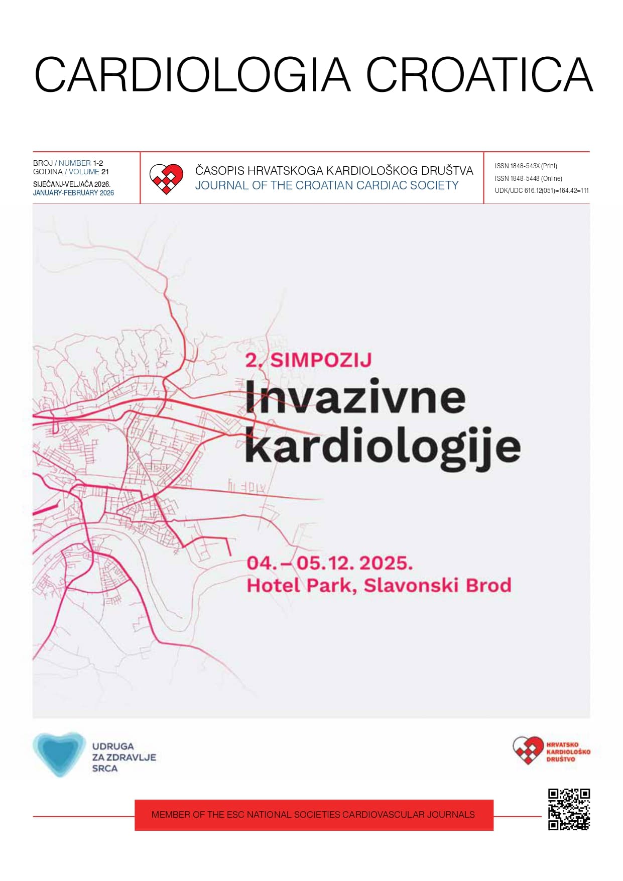
## Dear Colleagues, It is my great pleasure to welcome you to the 2nd Symposium on Invasive Cardiology in Slavonski Brod with international participation. Building on the success of our first meeting, which marked twenty years of invasive cardiology in Slavonski Brod, this year’s Symposium continues our shared commitment to professional growth, collaboration, and high-quality patient care. It reflects the ongoing development of our regional cardiac center and our dedication to applying advancements in modern cardiovascular medicine to our clinical practice. Our program will bring together experts in interventional cardiology, cardiac imaging, electrophysiology, and vascular medicine, as well as nurses and radiology engineers — all essential members of the heart team. The symposium encourages open exchange of knowledge and experience and promotes the multidisciplinary cooperation that defines good clinical practice. We are proud that, through teamwork and collaboration with national and international partners, Slavonski Brod has become recognized as a regional center where expertise, dedication, and education come together to improve patient outcomes. I would like to warmly thank all lecturers, participants, and colleagues who contributed to the organization this event, and our hospital administration for their continuous support and encouragement. I hope this year’s symposium will provide useful insights, new ideas, and strengthen the professional connections that move our field forward. Thank you for being part of it. Yours sincerely, Prim. dr. sc. Katica Cvitkušić Lukenda, dr. med. /
Kristina Vorkapić, Mario Špoljarić, Krešimir Gabaldo, Katica Cvitkušić Lukenda, Domagoj Mišković, Daniel Unić, Marijana Knežević Praveček
**Introduction:** Transcatheter aortic valve implantation (TAVI) in degenerated bioprosthetic valves, also known as valve-in-valve TAVI, is increasingly used in high-risk surgical patients. While commonly performed for structural valve deterioration causing stenosis, combined severe stenosis and regurgitation presents additional clinical and procedural challenges. (1-4) **Case report:** We report the case of a 69-year-old female with a history of arterial hypertension, hyperlipidemia, coronary artery disease, and previous surgical aortic valve replacement with a bioprosthetic valve and aortic root repair according to the Manougian technique 10 years prior. The patient presented with progressive dyspnea (NYHA Class III) and signs of congestive heart failure. Echocardiography revealed severe aortic stenosis (mean gradient 50 mmHg), severe central aortic regurgitation, preserved left ventricular ejection fraction (LVEF 50%), and significant left atrial enlargement. Given her prior cardiac surgery and high operative risk a multidisciplinary Heart Team recommended transfemoral valve-in-valve TAVI. Valve was implanted via transfemoral access under conscious sedation. Preprocedural CT imaging was used to confirm annular dimensions and coronary height. Deployment was guided by fluoroscopy and echocardiography. The valve was successfully positioned within the degenerated surgical bioprosthesis without complications. Post-procedure echocardiography confirmed appropriate valve positioning, no paravalvular leak, and complete resolution of aortic regurgitation. Peak velocity decreased to 3.5 m/s (mean gradient 26 mmHg). Left ventricular ejection fraction is mildly reduced at 40%. Before discharge, medical therapy for heart failure with reduced ejection fraction was optimized. **Conclusion:** Valve-in-valve TAVI is a safe and effective alternative for patients with failed surgical bioprosthetic valves and prohibitive surgical risk. This case highlights the feasibility of the procedure even in the presence of both severe stenosis and regurgitation, underlining the importance of detailed preprocedural imaging, careful valve sizing, and interdisciplinary planning. With appropriate patient selection, valve-in-valve TAVI offers excellent early outcomes and symptomatic relief in high-risk populations.
Marina Stanković, Alenka Tuličić-Mihelčić, Renata Valenčak, Saša Presežnik
Transcatheter aortic valve implantation (TAVI) is a minimally invasive procedure designed to treat severe aortic stenosis in patients who are considered high-risk candidates for conventional surgical valve replacement. Over the last decade, TAVI has become a widely accepted and routine cardiological intervention. The choice of anesthesia during TAVI is critical for patient safety and procedural success. Conscious sedation combined with local anesthesia is increasingly preferred over general anesthesia due to advantages such as shorter procedure times, faster patient recovery, and fewer anesthesia-related complications. At the General Hospital Slavonski Brod, TAVI procedures have been performed since early 2025 using conscious sedation alongside local anesthesia. In this multidisciplinary setting, perioperative nurses play a pivotal role throughout all stages of care. Their responsibilities include patient preparation, continuous monitoring of vital signs during the procedure, maintaining aseptic techniques, and ensuring patient comfort. Postoperatively, nurses oversee recovery, pain management, infection prevention, and patient education. A key focus of nursing care during TAVI is maintaining hemodynamic stability. Nurses monitor cardiovascular parameters closely, recognizing and responding promptly to any signs of hemodynamic instability or other complications. Effective analgesia and anxiety management are also essential nursing tasks, aimed at improving patient experience and facilitating smooth procedural flow. Moreover, perioperative nurses act as crucial communicators within the multidisciplinary team, ensuring seamless collaboration between cardiologists, anesthesiologists, and other healthcare professionals. This teamwork contributes to rapid identification and management of potential complications, ultimately enhancing patient safety and clinical outcomes. (1) This abstract highlight the vital nursing roles and responsibilities in the perioperative care of patients undergoing TAVI. Emphasizing patient safety, hemodynamic stability, pain control, and infection prevention, with perioperative nurses being key to the successful completion of the TAVI procedure.
Antonija Raguž, Marijana Knežević Praveček, Katica Cvitkušić Lukenda
**Introduction**: Mechanical heart valves are highly thrombogenic and require guided life-long anticoagulation. Their thrombosis is mainly subacute or chronic. Early diagnosis is crucial for preventing life threatening complications like valve obstruction, systemic embolization, cardiogenic shock. Treatment may vary according to patient´s clinical status, duration of symptoms, size of thrombus, severity of obstruction, anticoagulation status, operative and bleeding risk. Guidelines are given for optimizing anticoagulation, fibrinolysis and surgical procedures; transcatheter manipulation is an alternative method with promising mid-term results at experienced centers. (1-3) **Case report**: We present 74-year-old woman admitted with decompensated heart failure (NYHA IV) after unsuccessful diuretic escalation. She is a long-time diabetic, treated for atrial fibrillation, arterial hypertension and hyperlipidemia. 20 years ago, she underwent a procedure composed of ASD patch closure, mitral valve annuloplasty and single bypass grafting (VSM-OM). In 2017 two bare metal stents were implanted due to progressive coronary artery disease. In 2020. routine transthoracic ultrasound control revealed stenotic mitral valve with estimated MVA 1.3-1.4cm2, mild MR, heavily dilated left atrium and preserved left ventricular function. Coronary angiography showed diffuse multivessel disease, occluded bypass, strong collateral flow and LVEF 65% on ventriculography. On transesophageal echocardiography abundant fibrotic pannus ingrowth was visualized causing stenosis. Patient was subjected to mechanical mitral valve replacement. In the following years she experiences two ischemic cerebrovascular infarctions. Mitral prosthesis malfunction was not detected; INR was repeatedly 2-2.5 and carotid ultrasound showed intermediate stenosis. At current admission she was hypotensive with pulmonary edema. Acute coronary syndrome was excluded. Striking severe mitral stenosis was seen on transthoracic ultrasound, indicators of severe pulmonary hypertension, dilated RV with preserved contractility interrupting left ventricular systolic function, noticeable SEC in left atrium. Prosthesis thrombosis was suspected and transesophageal ultrasound was performed, thrombi in left atria were visualized with dense SEC, prosthesis itself was surrounded and invaded with pannus without obstructive thrombotic masses, severity of obstruction was confirmed (MVA 0.8cm2, mean gradient 22mmHg). Operative risk was deemed too high. She was discharged in NYHA III functional status with enoxaparin 1mg/kg subcutaneous twice daily, aspirin 75mg orally and optimal heart failure medical treatment. **Conclusion:** Our therapeutic option here is optimal anticoagulation. Heparin treatment is planned for 4 weeks, then reassessment to decide when to reintroduce warfarin with target INR >3 or to use t-PA if indicated.
Rea Levicki, Ivan Barišić, Jurica Petranović, Ile Raštegorac, Martina Lovrić Benčić
**Introduction:** Tetralogy of Fallot (TOF) includes: right ventricular (RV) hypertrophy, RV outflow tract obstruction, ventricular septal defect (VSD), and overriding aorta. (1) Long-term complications after complete TOF surgical repair, especially after transannular patch repairs, include; pulmonary regurgitation (PR), RV dysfunction, arrhythmias, and increased risk of sudden cardiac death. (2, 3) Optimal timing for severe PR intervention (implantation of a bioprosthetic pulmonary valve or transcatheter pulmonary valve implantation) is difficult to determine. (4) **Case report:** We present two patients with TOF, a 23-year-old man (M) and a 49-year-old woman (W), who were hospitalized for acute heart failure. In M total TOF correction was performed at the age of 3 with transannular patch repair of PA, postprocedural RVOT PG was 30 mmHg. W underwent TOF correction at the age of 5. Clinical examination in both patients confirmed a systolic murmur II/VI with punctum maximum above the pulmonary valve. A right bundle branch block was identified on the 12-lead electrocardiogram in both. Laboratory analysis demonstrated elevated levels of N-terminal pro–B-type natriuretic peptide (M 7677 pg/ml, W 968 pg/ml). Echocardiography (TTE) in M patient demonstrated a dilated, volume-overloaded RV with moderate RV base dilatation and significant RV apical dilatation, and mildly reduced RV systolic function (fractional area change 34%, TAPSE 14 mm, s’ 8.9 m/s), and a sever PR (PTH 79 ms, present PA reverse flow, regurgitation jet occupied 80% of the RVOT width, maxPG 30 mmHg, RV systolic pressure 35 mmHg), LV size and function were normal, without VSD (**Figures 1** and **2**Figure 2). TTE in W (**Figure 3**) showed a dilated, volume-overloaded RV with leftward shift of the interventricular septum in diastole and preserved RV systolic function (FAC 50%, TAPSE 20 mm), with significant PR (regurgitation jet occupied 80% of the RVOT width, VC 12 mm, maxPG 17 mmHg, RV systolic pressure 32 mmHg). They were both referred to tertiary adult congenital heart diseases (ACHD) center were the diagnosis was confirmed. W is a candidate for PV operation, and for M optimal medical treatment was recommended considering comorbidities. FIGURE 1. 23-year-old man: Volume-overloaded right ventricle with leftward shift of the interventricular septum in diastole. FIGURE 2. 23-year-old man: Color Doppler of the right ventricular outflow tract and branch pulmonary arteries in parasternal short axis view with the retrograde flow originating from the distal branch pulmonary arteries. FIGURE 3. 49-year-old woman: Color Doppler of the right ventricular outflow tract and branch pulmonary arteries in parasternal short axis view. **Conclusion:** These cases highlight the importance of echocardiographic detection of significant PR in TOF repair patients to prevent the development of RV dysfunction.
Anita Juričić, Matea Hiller, Ana Bukovac
**Introduction**: Transcatheter aortic valve implantation (TAVI) is a minimally invasive therapeutic procedure used to treat severe aortic stenosis in patients with high surgical risk. It involves the implantation of a bioprosthetic aortic valve via catheter, most commonly through the femoral artery, without the need for open-heart surgery or cardiopulmonary bypass. The decision for this procedure is made by a multidisciplinary team consisting of a cardiologist, cardiac surgeon, and anesthesiologist, based on comprehensive diagnostic evaluations (1). Common symptoms of aortic stenosis include shortness of breath, fatigue, palpitations, dizziness, and syncope. Major risk factors are advanced age and bicuspid aortic valve, alongside other contributing factors such as metabolic syndrome, diabetes, hypertension, smoking, hyperlipidemia, infections, and cardiovascular diseases. Diagnosis is based on echocardiography, ECG, laboratory tests, CT angiography, and coronary angiography (2). **Case report**: This case report presents an elderly female patient admitted to the cardiology department one day prior to the scheduled TAVI procedure. Upon admission, she reported no acute symptoms, was self-sufficient, but experienced exertional fatigue. Pre-procedural preparation included establishing venous access, collecting laboratory tests, performing ECG and cardiac ultrasound, and administering prophylactic antibiotic therapy. The TAVI procedure was performed via the femoral approach without complications. Post- intervention, the patient was transferred to the coronary care unit for continuous monitoring of cardiac rhythm, blood pressure, respiratory status, and femoral puncture site. Laboratory tests were conducted at 2-, 4-, and 8-hours post-procedure. The patient recovered well and was discharged on the third day after the intervention. **Conclusion**: Nurses play a critical role in the pre-, intra-, and post-procedural care of TAVI patients. Their responsibilities include monitoring vital signs, managing patient care, preventing complications, and educating patients (3). Patient education focuses on medication adherence, regular cardiology follow-ups, recognizing potential complications, and implementing lifestyle changes. By providing individualized care and support, nurses contribute significantly to the recovery process and long-term quality of life of patients undergoing TAVI (2).
Katica Cvitkušić Lukenda, Ivan Bitunjac, Domagoj Vučić, Marijana Knežević Praveček, Krešimir Gabaldo, Ivica Dunđer, Antonija Raguž, Domagoj Mišković, Zrinko Pešut, Josip Silović, Anto Lukenda, Vjekoslav Tomulić, Igor Medved, Jurica Juranić, Josip Samardžić
**Introduction:** The incidence of severe aortic stenosis is rising with population aging. Once symptomatic, the disease carries a poor prognosis if left untreated (1). Transcatheter aortic valve implantation (TAVI) provides a less invasive alternative to surgery, improving survival and quality of life in high-risk or inoperable patients. Common complications can be effectively managed in regional centers, while emergency surgery is rarely required (<0.5%) (2, 3). In 2025, a structured institutional TAVI program was initiated at the General Hospital “Dr Josip Benčević”, Slavonski Brod, in collaboration with the University Hospital Centre Rijeka, based on a multidisciplinary heart team approach and formal surgical backup. **Patients and Methods:** A retrospective, single-center analysis was performed using the institutional database. Demographic data, comorbidities, chronic therapy, echocardiographic parameters, procedural details, and in-hospital complications were descriptively analyzed. Functional status was assessed using the NYHA classification. **Results:** A total of twenty patients (mean age 79 years, 55% male) with severe aortic stenosis underwent TAVI procedures at our institution. All patients were symptomatic, with 65% presenting with advanced symptoms (NYHA class III–IV). The mean transvalvular pressure gradient was 55.4 mmHg, with an average LVEF of 57%. The most common comorbidities were hypertension (95%), hyperlipidemia (90%), and diabetes mellitus (50%), while prior myocardial infarction and coronary artery bypass graft were present in 30% and 15% of patients. Right femoral access was used in 85% of cases, with balloon predilatation in 80%. The 29 mm Evolut valve was the most implanted (75%), with all implantations achieving procedural success. Vascular access complications occurred in two patients, both requiring vascular surgery. One in-hospital death occurred 48 hours post-TAVI due to retroperitoneal hematoma in an 88-year-old woman with low BMI receiving chronic anticoagulation therapy. Conduction disturbances occurred in five patients, requiring permanent pacemaker implantation in two. One case of cardiac tamponade was successfully managed with pericardiocentesis. **Conclusion:** Our initial experience shows that TAVI without on-site cardiac surgery can be safely performed in well-prepared regional centers with multidisciplinary support and surgical backup. Such programs improve access and outcomes for high-risk patients, while continued team training and optimized patient selection remain essential.
Ivan Bitunjac, Katica Cvitkušić Lukenda
**Introduction**: Mechanical circulatory support (MCS) is increasingly used during high-risk percutaneous coronary interventions (HR-PCI), especially in patients with limited myocardial reserve and complex coronary anatomy (1). Pulsatile devices such as the iVAC 2L are typically positioned across the aortic valve for left ventricular (LV) unloading (2). However, in patients with mechanical aortic valves, this configuration is contraindicated (3). **Case report**: We report the case of a 73-year-old male with a history of complex cardiac surgery, including initial bioprosthetic aortic and mitral valve replacement followed by reoperation due to infective endocarditis, resulting in mechanical aortic valve implantation and prior coronary artery bypass grafting. He presented with exertional angina (CCS III). Coronary angiography revealed a severely calcified 70% stenosis of the left main coronary artery (LMCA), chronic total occlusion of the right coronary artery, and a patent previously implanted stent in the circumflex artery. All previous bypass grafts were found to be occluded. The patient was considered inoperable due to prohibitive surgical risk and referred for HR-PCI. To avoid crossing the mechanical prosthesis, the MCS catheter was positioned entirely within the ascending aorta, just above the valve (“aorta-only” configuration; **Figure 1**), with the aim of augmenting aortic pressure and maintaining coronary perfusion during the procedure. Intravascular lithotripsy and drug-eluting stent implantation of the LMCA were performed under intravascular ultrasound guidance. The procedure was successful and uneventful, with preserved renal function and resolution of anginal symptoms. FIGURE 1. iVAC 2L catheter positioned entirely within the ascending aorta, above the mechanical aortic valve, in an “aorta-only” configuration used to support high-risk percutaneous coronary intervention. **Conclusion**: This “aorta-only” approach represents a conceptual shift from traditional LV unloading toward pressure augmentation (2). It offers a novel and feasible strategy in anatomically and surgically complex cases where conventional support options are contraindicated (4). To our knowledge, this is the first report of mechanical circulatory support positioned exclusively in the ascending aorta during HR-PCI in a patient with a mechanical aortic valve.
Alenka Tuličić-Mihelčić, Renata Valenčak, Saša Presežnik, Marina Stanković
Atrial fibrillation (AF) is the most common cardiac arrhythmia in the general population. The global prevalence of AF is estimated to be between 2% and 4%, with a continued increase in the number of patients expected (1). AF is associated with increased overall mortality and morbidity, as well as a higher risk of stroke and heart failure (2). The gold standard in the treatment of AF is pulmonary vein isolation. At the General Hospital Slavonski Brod, the first pulmonary vein isolation using cryoablation was performed on February 13, 2019. Since then, a total of 186 procedures have been conducted. The procedure is performed by an interventional cardiologist/electrophysiologist. At the beginning of the procedure, local anesthetic is applied to both groin regions. The left femoral vein is punctured, and an introducer sheath is inserted through which a line for measuring activated clotting time, an invasive pressure monitoring system, and intracardiac echocardiography (ICE) are placed. Through the puncture of the right femoral vein, an introducer with an inserted needle for transseptal puncture is positioned, enabling access to the left atrium and pulmonary vein ostia. A cryoballoon is then inserted through the same introducer, and ablation of the pulmonary vein ostia is performed using liquid nitrogen. ICE is removed, and a decapolar electrophysiological catheter is placed to monitor the function of the phrenic nerve via electrical stimulation. If, at the end of the procedure, the patient is still in atrial fibrillation, synchronized cardioversion is performed (1). As with all invasive cardiology procedures, aseptic techniques, sterility protocols, hemodynamic monitoring, analgesia and sedation, coagulation time measurement, and fluoroscopy are all employed, alongside other standard procedural steps. The medical team includes two nurses and a radiology technologist. Potential complications include allergic reactions to the contrast agent, puncture site complications such as bleeding, hematoma, or infection, phrenic nerve injury, esophageal injury, cardiac perforation, pulmonary vein damage, or stroke. This is why the procedure requires thorough preparation and a well-trained, experienced team (2).
Jurica Petranović, Rea Levicki, Ivan Barišić, Ile Raštegorac, Richard Matasić
**Introduction:** Arrhythmogenic cardiomyopathy (ACM) is a hereditary heart muscle disease that can affect the isolated right ventricle (RV), both ventricles, or with isolated affection of the left ventricle (LV), in which the healthy myocardium is replaced by fatty and fibrous tissue. (1) It is inherited in an autosomal dominant manner. Genes coding for desmosomal and non-desmosomal proteins are most often mutated. Structural abnormality of the myocardium causes a disturbance in the electrical system of the heart, which manifests as ventricular tachycardia and subsequent sudden cardiac death (SCD). (2) Diagnosis is based on magnetic resonance imaging, echocardiography, and genetic testing. The main goal of treatment is the prevention of sudden cardiac death, with antiarrhythmic drugs to prevent the occurrence of malignant arrhythmias, and primary prevention of SCD by implanting an ICD. (3) **Case report:** 22-year-old patient was hospitalized due to recurrent syncope during exertion. Echocardiography showed a normal-sized left ventricle (EDD 48mm), normal-wall thickness (IVSd 9 mm), no segmental contractility loss, EFLV 60%, GLS: -18.2%, slightly decreased values for the basal part of the inferoseptal wall and the medial part of the lateral wall. Diastolic function was normal. E/e 7.6. The right ventricle was normal-sized and without trabeculation, with preserved systolic function. A 12-minute treadmill test was performed, with no discomfort during exercise, and no signs of ischemia on the electrocardiogram, but during recovery, persistent hemodynamically stable ventricular tachycardia with a frequency of 250/min was recorded (**Figure 1**), spontaneously converted to sinus rhythm, and short-term NSVTs were subsequently recorded (**Figure 2**). An MRI of the heart verifies the subepicardial zone of late post-contrast inhibition of the inferoseptal, inferior, and inferolateral walls in the basal and middle third, fulfilling the large structural MR criteria for left ventricular affection with ACM. He was medically treated with the maximum tolerated dose of propranolol. The patient was referred to a tertiary center for further treatment, and an extravascular cardioverter-defibrillator (Medtronic Aurora EV-ICD) was implanted. FIGURE 1. Ventricular tachycardia (ECG) on stress test (in recovery phase). FIGURE 2. Non sustained ventricular tachycardia on stress test (in late recovery phase). **Conclusion:** Special caution when examining patients with unexplained syncope during exertion is necessary in order not to overlook a possible malignant arrhythmia in undiagnosed ACM.
Zvonimir Katić, Matija Mlinar, Domagoj Kardum
Atrial fibrillation (AF) is the most prevalent cardiac arrhythmia, significantly affecting healthcare services (1). Pulmonary vein isolation (PVI) has emerged as the standard treatment for AF, with radiofrequency energy historically being the primary method employed. The second most common ablation technique has been cryoenergy, particularly using cryoballoon (CB) technology, which has demonstrated superiority over drug therapy in patients with paroxysmal atrial fibrillation (PAF) (2). Lately new energy source for PVI emerged. Pulsed field ablation (PFA) became very popular as a single shot method which is cardioselective (all tissue around heart is safe from damage such as the phrenic nerve and esophagus). As a very experienced center with single shot PVI method since introduction of PFA we performed more than 250 procedures. In the mixed paroxysmal and persistent population, up to 73.8% of patients remained free of AF recurrence in the 5-year follow-up, when accounting for redo procedures and AADs. Only 2.4% of patients experienced major complications of the ablation procedure, none with permanent sequelae. Now we have only short term follow up for patients with AF after PFA but results are promising. From 115 patients treated with PFA only 8 had reccurence of AF 6 months from first procedure.
Josip Ereiz, Ante Lisičić, Ana Jordan, Katica Cvitkušić-Lukenda
**Introduction**: Coronary artery (CA) injury is a rare complication following radiofrequency ablation (RFA) with the overall incidence of less than 0.1%. The risk of injury can be even higher (50%) if applying radiofrequency energy within 2mm of the CA. (1-3) **Case report**: The patient, a 36-year-old Caucasian female was referred for an electrophysiology (EP) study (April 2022) after narrow complex tachycardia had been diagnosed in the Emergency Department. The patient is a healthy adult with highly symptomatic episodes of palpitations which occurred daily. The EP study was conducted, confirming the diagnosis of typical atrioventricular nodal re-entrant tachycardia (AVNRT). After the radiofrequency energy was applied on the slow pathway, solely the atypical AVNRT was re-induced. The site of the earliest activation was located on the posteroseptal part of the right atrium, next to the ostium of the coronary sinus (CS). An additional ablation was performed and the tachycardia was terminated. After the procedure, a single episode of ventricular fibrillation was observed, necessitating prompt defibrillation (**Figure 1**). Once the return of the spontaneous circulation has been re-established, electrocardiogram demonstrated changes indicative of an acute myocardial infarction of the posterior region (**Figure 2**). The next step involved an urgent invasive coronary angiography, which confirmed acute occlusion of the posterolateral (PL) branch situated next to the ostium of the CS (**Figure 3**). Percutaneous transluminal coronary angioplasty was performed and re-established the blood flow through the PL branch, but on the follow-up angiogram, a localised contrast extravasation was observed. After a prolonged balloon inflation, normal flow through the PL was restored. Over the course of the remaining hospitalization, the patient was hemodynamically stable, without residual chest pain and without segmental wall motion abnormalities or pericardial effusion on echocardiogram. New arrhythmias were not detected. FIGURE 1. Ventricular fibrillation after radiofrequency ablation. FIGURE 2. Acute posterior myocardial infarction after radiofrequency ablation. FIGURE 3. Coronary angiography, occluded posterolateral branch. **Conclusion**: Even though the RFA is highly effective, caution is necessary when applying energy within the CS due to its close anatomical correlation with PL branch. Performing coronary angiography prior to the energy delivery may aid in the prevention of CA injury.
Ivica Dunđer, Katica Cvitkušić Lukenda, Ivan Bitunjac, Marijana Knežević Praveček, Krešimir Gabaldo, Domagoj Mišković, Domagoj Vučić
**Introduction**: Complete atrioventricular (AV) block is a life-threatening condition characterized by a partial or complete interruption of impulse conduction from the atria to the ventricles. The most common causes are fibrosis and sclerosis of the conduction system, ischemic heart disease, and electrolyte imbalance (1). **Case report**: 85-year-old patient was admitted via the Emergency Department due to recurrent episodes of loss of consciousness, with ECG showing complete AV block and a ventricular rate of 28 bpm (**Figure 1**). Multiple comorbidities were present, including type 2 diabetes mellitus with complications, arterial hypertension, and a history of stroke 30 years ago, with resulting right-sided hemiparesis and aphasia. Temporary external pacing electrodes were promptly placed, and upon admission to the Coronary Care Unit, a temporary pacemaker was inserted via the right jugular vein, after which the patient was stabilized. On the following day, a permanent dual-chamber pacemaker was implanted via the cephalic vein, with excellent stimulation parameters (**Figures 2** and **3**Figure 3). On the third day post-implantation, telemetry recorded an asystolic episode with cardiorespiratory arrest and loss of pacemaker output, prompting resuscitation and re-initiation of external pacing. A temporary pacemaker was reinserted, and the revision of the previously implanted pacemaker was performed. Radiographic imaging revealed no displacement of the electrode, with identical positioning and normal electrode impedance. The ventricular electrode failed to transmit impulses even when connected to the programmer. Therefore, a new ventricular electrode was implanted via the left subclavian vein (**Figure 4**). FIGURE 1. 12-lead electrocardiogram on admission. FIGURE 2. 12-lead electrocardiogram after pacemaker implantation. FIGURE 3. Chest X-ray at initial implantation. FIGURE 4. Chest X-ray after implantation of the new ventricular electrode. **Conclusion:** Dysfunction of the ventricular electrode may have been due to a microfracture of the electrode, which was not visible radiographically, or a microtear into fibrotic tissue (2).
Ivica Dunđer, Katica Cvitkušić Lukenda, Marijana Knežević Praveček, Domagoj Mišković, Sanja Radanac, Krešimir Gabaldo
**Introduction:** Optimal medical management of peripheral arterial disease (PAD) involves a combination of lifestyle modifications, pharmacologic therapy, and risk factor control to reduce symptoms, improve functional status, and prevent cardiovascular events (1). Lifestyle modification remains the cornerstone of PAD management and includes smoking cessation, supervised exercise therapy (SET), and dietary changes. Pharmacologic therapy consists of antiplatelet agents (aspirin 75–325 mg/day or clopidogrel 75 mg/day), and in patients at high ischemic risk, low-dose rivaroxaban (2.5 mg twice daily) should be added. Statin therapy is recommended for all patients, with a target LDL level 4.1 mmol/L, referred to our institution for intermittent claudication of the right leg after 100 meters. The initial ankle-brachial index (ABI) on the right was 0.65, and MSCT angiography showed a long occlusion (>150 mm) of the superficial femoral artery (SFA). A multidisciplinary team initially opted for surgical treatment with femoral-popliteal bypass. He was discharged on postoperative day 7 but readmitted 14 days later due to acute graft failure, requiring repeat surgery with Fogarty thrombectomy. Despite dual therapy (aspirin + warfarin), control Doppler imaging showed bypass occlusion. Endovascular recanalization was attempted via antegrade-retrograde access but was unsuccessful. The patient was prescribed SET and optimized medical therapy with aspirin + rivaroxaban (2.5 mg twice a day), high-dose statin (atorvastatin/ezetimibe), and perindopril/amlodipine. He also successfully quit smoking. At 3-month follow-up, he reported improved walking distance (up to 3 km) and ABI improvement to 0.86. **Conclusion:** Optimal medical therapy, combined with lifestyle modification—especially SET—is essential in PAD management. Revascularization should be reserved for cases where medical therapy fails or in critical limb ischemia.
Ivica Dunđer, Tea Cvetko, Sergej Nadalin, Ines Rajkovača Latić, Vesna Zarezovski Stilin, Katica Cvitkušić Lukenda, Zvonimir Bosnić, Domagoj Vučić
**Introduction**: Myocardial infarction with non-obstructive coronary arteries (MINOCA) is a heterogeneous clinical entity that accounts for approximately 5–10% of all acute myocardial infarction (AMI) cases. Among its underlying mechanisms, spontaneous coronary artery dissection (SCAD) represents an increasingly recognized cause, particularly in young women without traditional cardiovascular risk factors. Prompt diagnosis is crucial, as management strategies differ from those for atherosclerotic coronary disease. (1-3) We present a case of a young female patient with MINOCA due to spontaneous dissection of the left anterior descending (LAD) artery. **Case report**: 44-year-old woman was hospitalized due to chest pain, elevated high-sensitivity troponin levels, and a normal ECG, presenting with clinical features consistent with NSTEMI. Transthoracic echocardiography revealed preserved left ventricular systolic function, no significant valvular or structural abnormalities, and a subtle segmental wall motion abnormality of the apical region consistent with hypokinesia. Coronary angiography demonstrated a type 2 spontaneous dissection of the distal left anterior descending (LAD) artery (**Figure 1**). The patient was treated with dual antiplatelet therapy (aspirin and clopidogrel), a statin, an ACE inhibitor, and other supportive therapy, resulting in normalization of cardiac enzyme levels. Twenty-four hours after discharge, she was re-admitted due to recurrent chest pain and a renewed increase in cardiac biomarkers; repeat coronary angiography showed no progression of the LAD dissection. After an additional nine days of conservative in-hospital management, the patient was discharged home. Due to the persistent apical hypokinesia, cardiac magnetic resonance imaging (CMR) was performed, demonstrating an area of acute myocardial injury in the apical region of the left ventricle (**Figure 2**). However, because of ongoing myocardial edema, the extent of fibrosis could not be reliably quantified. Two months after the initial event, repeat coronary angiography demonstrated recovery of flow in the distal LAD segment (**Figure 3**). Clinically, the patient remained asymptomatic, and follow-up echocardiography showed no significant changes compared to the prior examination. FIGURE 1. Angiographic view of a type 2 dissection in the distal left anterior descending artery. FIGURE 2. Cardiac magnetic resonance imaging showing the area of acute myocardial injury and edema with late gadolinium enhancement. FIGURE 3. Angiographic image showing healing of the distal left anterior descending artery dissection. **Conclusion**: Spontaneous coronary artery dissection is an important and often underrecognized cause of MINOCA, predominantly affecting young to middle-aged women without conventional cardiovascular risk factors. Early identification through coronary angiography and, when necessary, intracoronary imaging or CMR is crucial for accurate diagnosis and appropriate management. Conservative treatment is generally preferred in hemodynamically stable patients, as most dissections heal spontaneously over time.
Alenka Tuličić-Mihelčić, Renata Valenčak, Saša Presežnik, Marina Stanković, Mato Čizmić, Katica Cvitkušić Lukenda
**Introduction**: The purpose of this case report is to demonstrate the complexity of percutaneous coronary interventions, the role of the Impella heart pump in providing adequate hemodynamic support, and the importance of the preparedness and education of all team members to successfully perform such high-risk and demanding procedures (1). **Case report**: 77-year-old patient with significant comorbidities and multiple malignancies in remission underwent coronary angiography, which revealed multivessel disease – a chronic total occlusion (CTO) of the right coronary artery, 70% stenosis of the left main (LM) coronary artery, 90% stenosis of the left anterior descending artery (LAD), and diffuse changes in all coronary vessels. Due to significant comorbidities and extremely high surgical risk, the cardiac surgeon did not recommend surgical revascularization. The interventional cardiologists in our department decided to proceed with percutaneous coronary intervention (PCI) supported by the Impella system, as the patient already showed signs of hemodynamic instability during the angiography itself. Since the patient also had peripheral vascular disease, percutaneous transluminal angioplasty (PTA) and balloon dilatation of the femoral artery were first performed to allow placement of the Impella CP system into the left ventricle. Lesion preparation was carried out using rotational atherectomy, cutting and scoring balloons, and Shockwave intravascular lithotripsy (IVL). Intravascular ultrasound (IVUS) confirmed adequate lesion preparation, and two stents were implanted – distally 3.0x28 mm and proximally 3.5x44 mm – optimized by post-dilatation. Good stent expansion and apposition were confirmed by final IVUS, after which the Impella system was removed. Hemostasis was achieved using two Prostyle and one AngioSeal vascular closure device. **Conclusion**: The patient was discharged home with stable vital signs, hemodynamically and cardiopulmonary compensated. Four months later, during a follow-up examination, he reported feeling well, without chest pain, and with optimal vital parameters.
Josip Silović, Domagoj Mišković, Katica Cvitkušić Lukenda, Krešimir Gabaldo, Marijana Knežević Praveček
**Introduction**: Mechanical complications of myocardial infarction (MI) represent rare but potentially fatal sequelae of acute MI that, despite advanced reperfusion therapies, maintain extremely high mortality rates (1, 2). These complications include left ventricular free wall rupture, interventricular septal rupture, papillary muscle rupture, and cardiac tamponade. Quick diagnosis is of utmost importance and echocardiography is the most available method for establishing it (3, 4). Aim: To analyze demographic characteristics, types of complications, management approaches, and short-term outcomes of patients with mechanical complications of MI treated at General Hospital Slavonski Brod during a five-year period. **Patients and Methods**: All patients with mechanical complications of MI hospitalized between January 2020 and October 2025 were retrospectively analyzed. Data were collected from the hospital information system and cardiology department database. Demographic characteristics, types of complications, infarct localization, culprit coronary vessels, management approaches, and short-term outcomes were analyzed. All diagnoses were confirmed by echocardiography. **Results**: A total of 11 patients were identified with a mean age of 73.8 years (range 61-88 years) and female predominance (54.5%). The most common complication was left ventricular free wall rupture (54.5%), followed by interventricular septal rupture (27.3%) and cardiac tamponade (18.2%). The right coronary artery was the most common culprit vessel (45.5%), while inferoposterior localization predominated in 36.4% of cases. Surgical management was applied in 54.5% of patients. Two cases had spontaneous cessation of bleeding without the need for surgical management: one case of cardiac tamponade where spontaneous cessation occurred after pericardiocentesis without a clear site of extravasation, and one case of free wall rupture with pseudoaneurysm formation. In one patient, percutaneous occlusion of the extravasation site with coils was successfully performed. Overall mortality was 30.0% (**Table 1**, **Figures 1** and **2**Figure 2). ### TABLE 1: Characteristics of patients with mechanical complications of myocardial infarction (General Hospital Slavonski Brod, 2020-2025). | **Patient** | **Age (years)** | **Sex** | **Complication Type** | **Culprit Vessel** | **Infarct Location** | **Management** | **Outcome** | | --- | --- | --- | --- | --- | --- | --- | --- | | 1 | 71 | F | Free wall rupture | RCA | Inferoposterior | Surgical | Survived | | 2 | 72 | F | Free wall rupture | RCA | Unspecified | Pericardiocentesis + coil occlusion | Survived | | 3 | 67 | M | Free wall rupture | RCA | Inferoposterior | Surgical (Patch repair) | Survived | | 4 | 64 | F | Cardiac tamponade | LCX | Lateral | Conservative | Deceased | | 5 | 74 | M | Free wall rupture | LAD | Anteroseptal | Conservative (pseudoaneurysm formation) | Survived | | 6 | 72 | M | Free wall rupture | LCX | Inferoposterior | Surgical | Survived | | 7 | 84 | F | Cardiac tamponade | Multiple culprits | Inferolateral | PCI + pericardiocentesis | Survived | | 8 | 68 | F | Free wall rupture | RIM | Inferolateral | Surgical | Deceased | | 9 | 60 | M | Septal rupture | RCA | Inferoposterior | Surgical (VSD patch) | Survived | | 10 | 87 | F | Septal rupture | LAD | Anteroseptal | Surgical | Deceased | | 11 | 68 | M | Septal rupture | RCA | Inferior | PCI + surgical | Unknown | [†] F – female; M – male; RCA – right coronary artery; LCx – left circumflex artery; LAD – Left anterior descending artery; PCI – percutaneous coronary intervention FIGURE 1. Survival outcomes by complication type. FIGURE 2. Number of patients by culprit vessel. **Conclusion**: Mechanical complications of MI occur more frequently in elderly women. Free wall rupture was the most common complication in our series. Mortality of 30.0% is significantly lower than published data, likely due to rapid echocardiographic diagnosis, 24/7 interventional cardiology availability, and established protocols for multidisciplinary management. Cases with spontaneous cessation of bleeding were identified where surgical management was not required, emphasizing the importance of individualized assessment of each patient. Surgical intervention remains the method of choice for definitive treatment whenever feasible, while percutaneous techniques are used selectively. RCA – right coronary artery; LCx – left circumflex artey; LAD – left anterior descending artery; RIM – ramus intermedius.
Zrinko Pešut, Ivan Bitunjac, Domagoj Mišković, Ivica Dunđer, Nikola Gotovac, Blaženka Miškić, Katica Cvitkušić Lukenda
**Introduction:** A coronary artery fistula (CAF) represents an abnormal connection between a coronary artery and a cardiac chamber or great vessel, either congenital or acquired. The incidence of CAF on coronary angiography (CAG) is 0.2–0.6%, most commonly originating from the right coronary artery (RCA, 50–60%), left anterior descending (LAD, 25–42%), or circumflex artery (ACx, 18%). Small CAFs are often asymptomatic, while larger fistulas may produce myocardial steal, ischemia, or heart failure. Chronic high-flow fistulas can cause aneurysmal dilatation or thrombosis, requiring percutaneous transcatheter closure (TCC) or surgical ligation. (1-4) **Case report:** 58-year-old man presented with non-ST-elevation myocardial infarction (NSTEMI). CAG revealed subocclusive stenosis of the proximal LAD, 60–70% stenosis of the distal ACx and obtuse marginal (OM) 1, and no significant RCA stenosis. Percutaneous coronary intervention (PCI) with two drug-eluting stents (DES) was performed in the LAD. Incidentally, CAFs from the proximal LAD and RCA to the pulmonary artery (PA) were suspected on angiography (**Figure 1****).** Multislice computed tomography coronary angiography (MSCT) demonstrated a significant RCA-to-right atrium (RA) fistula located between the aorta and PA, forming a small vascular conglomerate adjacent to the RA (**Figures 2** and **3**Figure 3). Cardiac magnetic resonance imaging (MRI) was recommended for cardiac pulmonary artery flow (CPAF) and Qp:Qs ratio to assess shunt volume. The need for percutaneous closure will be evaluated based on these findings. FIGURE 1. Coronary angiogram of the coronary artery fistula originating from the proximal left anterior descending artery. FIGURE 2. Multislice computed tomography coronarography image of the coronary artery fistula originating from the proximal right coronary artery, located between the aorta and the pulmonary artery. FIGURE 3. Multislice computed tomography coronarography imaging of the coronary artery fistula lying between the aorta and the pulmonary artery and continuing into a vascular conglomerate. **Conclusion:** Coronary artery fistulas, although rare, may have serious clinical implications. When detected by angiography, complementary imaging with CT or MRI helps define anatomy and quantify shunt flow. In significant cases, percutaneous TCC should be considered to prevent ischemia, heart failure, or aneurysmal complications.
Domagoj Mišković, Krešimir Gabaldo, Josip Silović, Ivica Dunđer, Zrinko Pešut, Antonija Raguž, Ivan Bitunjac, Domagoj Vučić, Marijana Knežević Praveček, Katica Cvitkušić Lukenda
**Introduction:** Coronary artery calcification (CAC) complicates percutaneous coronary intervention (PCI) by impairing stent delivery and expansion (1). Currently, in the battle against CAC, several tools are available, which can be divided into modified balloons, atheroablative technologies, and intravascular lithotripsy (IVL) (2). Rotational atherectomy (RA) facilitates lesion modification but carries specific risks. RA of o specific lesions: ostial right coronary artery (RCA), ostial left circumflex (LCX) with substantial bending, unprotected left main, diffuse long lesions can be very challenging. Since clinical outcomes of RCA ostial lesions have been unsatisfactory for decades, RA has been a good indication for RCA ostial lesions with severe calcification (3). **Case report:** Seventy-eight years old years female patient was admitted because of unstable angina pectoris. Urgent angiography showed severe calcified ostial RCA lesion. The first attempt at PCI was unsuccessful due to the lack of coaxial positioning of the guiding catheter. Finally, in a second attempt, a successful PCI RCA with single stent implantation was performed. The use of a long, 7-French femoral sheath (Destination Guiding Sheath, Terumo) was crucial for successful maneuvering and coaxial positioning of the guiding catheter. Lesion preparation was done with a 1.25 burr and non-compliant balloons (**Figures 1-3**Figure 2Figure 3). FIGURE 1. Calcified ostial right coronary artery lesion. FIGURE 2. The passage of the burr. FIGURE 3. Final angiogram after stent implantation. **Conclusion:** Reason why RA for RCA ostial lesions are difficult are impossibility of insertion guiding catheter to RCA coaxially and difficulties keeping catheter coaxial to RCA in aorta. Also, coaxial cannot be confirmed by left oblique view which is the standard projection for RA of RCA. There is additional risk of cerebral infarction, so it is important to use small burrs to minimize the size of debris. Although it is demanding and challenging, RA has been a good indication for RCA ostial lesions with severe calcification (3).
Marijana Knežević Praveček, Krešimir Gabaldo, Domagoj Mišković, Ivan Bitunjac, Ivica Dunđer, Antonija Raguž, Josip Silović, Zrinko Pešut, Renata Valenčak, Katica Cvitkušić Lukenda
Drug-eluting stents (DES) remain the standard of care in contemporary percutaneous coronary intervention (PCI). However, their permanent metallic scaffolding may pose long-term risks, including late stent thrombosis and in-stent restenosis (ISR). In response, “leave nothing behind” strategies particularly drug-coated balloons (DCB) are increasingly being adopted as a viable alternative. DCBs have demonstrated reliable clinical efficacy, especially in the treatment of ISR, and currently represent the only stent-free technology routinely used in PCI practice. Beyond ISR, their potential role in the treatment of de novo lesions is increasingly supported by emerging evidence. (1, 2) At the General Hospital in Slavonski Brod, Croatia, DCBs have been in use since 2010. Between October 2020 and October 2025, a total of 552 DCB procedures were performed out of 3.449 PCI procedures (16%). Annual use has remained stable and targeted. This rational and individualized application mirrors global trends, where DCB usage accounts for 6% to 16.5% of all PCI procedures in some regions and even higher in specialized centers. The 2018 European Society of Cardiology (ESC) guidelines on myocardial revascularization issued a class IA recommendation for the use of DCBs in the treatment of ISR. In contrast, the 2024 ESC guidelines for chronic coronary syndromes now recommend DES over DCBs in patients with DES-ISR (also class IA). In our center, we aim to make rational and individualized therapeutic decisions. Accordingly, in the treatment of ISR particularly in cases with multiple stent layers, persistent stent underexpansion, bifurcation lesions, or increased bleeding risk we consider DCBs a more acceptable and appropriate therapeutic option. It is important to emphasize that we do not view DCBs as a replacement for DES, but rather as a complementary technology particularly valuable for reducing the “metal burden” in complex or high-risk patients. The concept of “hybrid” PCI strategies (DCB + DES) is gaining momentum and supports an individualized approach. Our experience confirms that when used thoughtfully, DCB angioplasty is a safe, effective, and sustainable revascularization strategy that deserves a more prominent role in future PCI paradigms.
Vedrana Baraban, Nika Srb, Ninoslava Vonić
The 2019 ESC guidelines first introduced chronic coronary syndrome (CCS) as a more comprehensive, diagnostically and therapeutically demanding clinical entity than the previous term stable angina pectoris. It includes the clinical presentation of coronary artery disease (CAD) that occurs during a stable period of coronary disease, after or preceding acute coronary syndrome. CCS encompasses various clinical presentations of stable ischemic heart disease. Thanks to new insights into the pathophysiological mechanism of ischemia, CCS is not associated exclusively with obstructive atherosclerotic disease of the epicardial coronary arteries, but also with functional and structural disorders of the microcirculation, vasospasm and endothelial dysfunction, i.e. anginal disorders within the framework of angina/ischemia with no obstructive coronary artery disease (ANOCA/INOCA). Men have about 50% higher risk of developing CCS, but ANOCA/INOCA clinical entities are higher in women. Along with atherosclerotic vascular changes (risk factors: diabetes, arterial hypertension, obesity, dyslipidemia), nonvascular causes of CSS are increasingly being investigated in the pathogenesis of myocardial ischemia, such as myocardial metabolic disorders, coronary flow changes due to platelet activation, microembolization, and endothelial inflammation. According to the 2024 ESC guidelines, there are 5 categories of CSS: patients with proven coronary artery disease and anginal disorders during exercise, patients with anginal disorders (vasospasm or microcirculatory disease) but without obstructive coronary disease (ANOCA/INOCA), patients with stable symptoms after ACS or revascularization, patients with stable symptoms and heart failure of ischemic or cardiometabolic etiology, and the asymptomatic category in which CAD was detected by invasive or noninvasive imaging methods, due to other indications. It is crucial to identify and effectively manage all aspects of Chronic Stable Syndrome (CSS) promptly, as they often go undiagnosed despite causing significant morbidity, reduced quality of life, and cardiovascular complications such as arrhythmias, valvular diseases, heart failure, and major adverse cardiac events (MACE). Although there has been notable progress in understanding the mechanisms behind CSS and in current treatments—including lifestyle modifications—a considerable number of patients, particularly those asymptomatic, remain undetected. Additionally, further clinical research is needed to refine antianginal therapies and invasive coronary procedures, aiming for comprehensive and long-term stabilization of CSS. (1-4)
Nikolina Bukal Ćaleta, Marijana Kovačević, Ivana Stanušić, Iva Dumančić, Josipa Meter, Ana Kovačević, Maja Franić
Both guidelines; ESC 2024 and ACC/AHA 2025 confirming true resistant hypertension as the official values of blood pressure (BP) >140/90 mmHg despite 3 or more BP-lowering medications at maximally tolerated doses including diuretic, by excluding pseudo-resistance (improper measurement, white coat effect, poor adherence, suboptimal regimen/dosing) and causes of secondary hypertension (primary aldosteronism, renal artery stenosis, obstructive sleep apnea, drug/substance contributors, thyroid disease, Cushing, chronic kidney disease etc.). New ACC/AHA guidance expands recommendation to screen more broadly for primary aldosteronism in patients with resistant hypertension (including many without hypokalemia). Out-of-office BP measurement (HBPM/ABPM) is strongly recommended. Both favor the usual 3-drug foundation (ACEi/ARB + calcium channel blocker +thiazide-like diuretic). If blood pressure remains uncontrolled on these, mineralocorticoid receptor antagonist (MRA) spironolactone (or eplerenone if spironolactone not tolerated), is the preferred fourth-line agent in many patients with cautions for renal function and hyperkalemia (ESC Class IIa). If spironolactone is not tolerated, alternatives include amiloride or a beta-blocker depending on indications. After confirming true resistant hypertension, excluding secondary causes, optimizing the therapy and adding spironolactone as preferred 4th-line, refer to specialist centers for complex causes and consider renal denervation (RDN) only as a specialized shared-decision option after thorough evaluation. ESC 2024 now permits consideration of renal denervation for selected patients with resistant uncontrolled hypertension after shared decision-making and specialist assessment and it is not recommended in severe CKD (eGFR <40) or secondary hypertension. (Class IIb) (1) ACC/AHA materials discuss RDN as an emerging option but the 2025 ACC/AHA update centers on standardized pharmacologic and diagnostic pathways in primary care. (2)
Haris Porobić
In interventional procedures, the clarity and accuracy of imaging are crucial for successful outcomes. Interventional procedures, including angiography, stent placement, and minimally invasive surgeries, rely heavily on high-quality imaging. Diascopy, a technique involving the manipulation of imaging parameters such as brightness, contrast, and zoom, significantly influences image quality. Image quality in interventional imaging depends on multiple factors, including Resolution, Signal-to-noise ratio (SNR), Contrast-to-noise ratio (CNR), Brightness and contrast settings, Geometric magnification or zoom, Image filtering and processing algorithms Diascopy protocols are predefined for certain procedures and patient groups where diascopy parameters are automatically selected. In 96% of cases, no adjustment is required. In 4% of patients, it is not possible to achieve satisfactory image quality, display of fine details or implant structures. Although the percentage of such cases is small, these patients are life-threatening and the possibility of image optimization is of great importance. (1) Hypothesis: Manual adjustment of diascopy parameters can significantly affect image quality and treatment outcome. **Patients and Methods:** The study was conducted at the Department of Invasive Cardiology, University Clinical Center of Sarajevo (KCUS), from February 1, 2022, to July 31, 2024. The study included patients of both sexes and different age groups. The study included hemodynamically stable patients, patients with myocardial infarction who underwent stenting of a lesion in the coronary blood vessels. Hemodynamically unstable patients, minors, and unconscious patients were not included in the research. The procedures were performed in the cardiac catheterization room on a TOSHIBA INFINIX diascopy device. Toshiba Infinix can manually adjust all diascopy parameters (kV, mA, matrix, pulse width, number of frames, three types of collimations), which return to system-defined values after the procedure is complete. It is this technical capability that made this type of research and proof of our hypothesis possible. The diagnostic acceptability of the image was evaluated by three examiners with grades from 1 to 5: [list-style:none] 1. 1 - non-diagnostic (means that the obtained structures cannot be analyzed), 2. 2 - unclear anatomical details with worsening image quality, 3. 3 - clearly increased clouding of the image, which is still not reflected in the diagnostic quality of the image, 4. 4 - clearly visible anatomical details with medium blurring of the image, 5. 5- clear delineation of small structures with clearly visible anatomical details and sharply delineated stent structures **Results and Conclusion:** In the control group of 800 participants, selected based on the study inclusion criteria, the image quality was evaluated. There were 531 (66%) males and 269 (34%) females. According to the subjective assessment of image quality with implanted stents, the evaluators rated the images on a scale from 1 to 5. The quality scores of two groups of images were compared: with (90 kV and 900 mA) and (150 kV and 600 mA), where a statistically significant difference was found in the image quality score of the two groups depending on the electrical potential (p<0.05). The quality ratings of two groups of images with different frame rates were compared, where no statistically significant difference was found in the image quality ratings of the two groups depending on the frame rate (p=0.767), however, the radiation dose increased by five times.
Ivana Simić, Barica Stanić
Cardiovascular diseases are the leading cause of mortality worldwide, and their timely recognition and monitoring are crucial for reducing complications and improving patients’ quality of life. Traditional monitoring of cardiac patients relies on hospital telemetry and periodic examinations. This type of monitoring is feasible only for hospitalized patients and ceases after discharge. However, the advancement of digital technologies has opened new possibilities. Modern tools such as smartwatches are now capable of tracking heart rate, rhythm, ECG recordings, oxygen saturation, and activity levels. Validation studies demonstrate high accuracy in detecting atrial fibrillation, comparable to traditional methods (1, 2). The introduction of artificial intelligence further enhances the efficiency of algorithms for arrhythmia detection (2). Mobile applications constitute a significant part of the digital ecosystem in cardiology care. Some connect with smart devices to automatically collect vital data, while others enable patients to manually input data. The implementation of telemedicine has further improved access to care, especially in remote areas. Telemedicine platforms enable remote consultations, visual patient monitoring, and integration of data from wearable devices. Meta-analyses show that this approach reduces mortality rates and the frequency of rehospitalizations (3). New generations of integrated systems allow automatic analysis of vital parameters and the sending of alerts if deviations are detected (e.g., weight gain in heart failure patients) (4). Digital monitoring of cardiology patients represents a significant advancement in contemporary cardiac care. This approach, however, carries challenges such as digital illiteracy, device unreliability, and personal data protection. It is important to note that technology does not replace healthcare professionals but complements them, enabling better disease management. The nurse plays a crucial role in implementing digital monitoring. She educates the patient on proper device usage, analyzes data, recognizes changes, and coordinates further interventions in collaboration with the interdisciplinary team. They facilitate education, monitoring, analysis, and human interaction. Therefore, it is crucial to invest in the education and training of healthcare professionals, the standardization of medical devices, and ensuring equitable access to technology for all patients, irrespective of their age or level of digital literacy.