Cardiologia Croatica
Journal Research Assistant
Journal Research Assistant
Journal Research Assistant
Journal Research Assistant
Ivica Benko, Mateja Lovrić, Marina Žanić, Marina Budetić, Nikolina Slamek, Mirela Adamović, Ružica Lovrić, Ana-Maria Goronić, Mario Tomašević, Ivan Horvat
**Introduction**: Ventricular fibrillation (VF) storm is a life-threatening condition characterized by recurrent malignant ventricular arrhythmias often refractory to antiarrhythmic therapy. Despite optimal revascularization and pharmacological management, this entity remains a major therapeutic challenge requiring urgent multidisciplinary action (1). **Case report**: We present a 58-year-old male with recent anterior ST-elevation myocardial infarction treated with percutaneous coronary intervention of the left anterior descending artery, admitted after out-of-hospital cardiac arrest due to ventricular fibrillation (VF). During the following days, recurrent VF storm occurred, necessitating >150 defibrillations despite intravenous amiodarone, lidocaine, beta-blockers and electrolyte optimization. The patient required deep sedation, mechanical ventilation and hemodynamic support. Urgent electrophysiological study with 3D electroanatomical and intracardiac echocardiography-guided mapping revealed apical scar substrate. Catheter ablation of triggering premature ventricular complexes was performed with transient rhythm stabilization. Subsequent course was complicated by sepsis, acute renal failure requiring dialysis and multi-organ dysfunction. After prolonged intensive care, tracheostomy and gradual recovery, left ventricular ejection fraction improved from 20% to 35%. The patient was weaned from mechanical ventilation and transferred to a regional hospital for further treatment. **Conclusion**: VF storm represents an extreme medical emergency with high mortality. Successful management requires rapid recognition, advanced interventional strategies such as catheter ablation, and coordinated intensive care. This case emphasizes the crucial role of highly trained nurses across the continuum of care, from intensive cardiac units to electrophysiology laboratories, whose expertise and timely interventions are essential in stabilizing patients during the most stressful and life-threatening arrhythmic scenarios.
Magdalena Drljačić, Ivica Benko, Ivona Filipović, Ivana Šmuc, Mateja Lovrić, Marina Žanić, Nikolina Slamek, Mirela Adamović, Senka Pejković
**Introduction:** Wide QRS tachycardia represents one of the most challenging findings in cardiology, as it can correspond to several underlying mechanisms: ventricular tachycardia (VT), supraventricular tachycardia (SVT) with aberrant conduction, pre-excitation syndromes, or drug/electrolyte-induced conduction disturbances. Since inappropriate classification may result in delayed or inadequate treatment, the general rule in doubtful cases is to treat the arrhythmia as VT until proven otherwise (1-3). **Case report:** 75-year-old female with a history of hypertrophic cardiomyopathy, Wolf-Parkinson-White syndrome, and type 2 diabetes was admitted following a 24-hour Holter ECG that revealed multiple sustained wide QRS tachycardias, symptomatic with weakness and dizziness, but without syncope. The Holter recorded sinus rhythm as the baseline, with 11,811 ventricular and 1,228 supraventricular extrasystoles. During symptomatic episodes, wide QRS tachycardia was documented, but it was unclear whether the mechanism was ventricular or supraventricular with aberrancy. The patient was referred for urgent hospitalization, and after diagnostic work-up including echocardiography and laboratory tests, she underwent an electrophysiological study. A sustained clinical tachycardia was induced, consistent with orthodromic AV reentrant tachycardia via a left lateral accessory pathway. Radiofrequency ablation was performed successfully with subsequent loss of conduction through the pathway. Post-ablation, tachycardia was no longer inducible and telemetry remained stable throughout hospitalization. **Conclusion.** This case emphasizes the complexity of interpreting wide QRS tachycardias in Holter analysis and the need to approach uncertain cases as potential VT. It also highlights the importance of comprehensive clinical education of nurses involved in Holter monitoring, extending beyond ECG curve recognition to an integrated understanding of arrhythmia mechanisms, clinical context, and patient safety.
Sara Milanović Litre, Anđelo Vukić
**Introduction:** Left bundle branch area pacing (LBBAP) is increasingly used to treat bradycardia and deliver cardiac resynchronization therapy (CRT) (1). During LBBAP, QRS morphology in lead V1 is characterized by a terminal R wave, known as the right bundle branch block (RBBB) pattern; however, different QRS complexes have been described (2). This study aimed to analyze QRS morphology in lead V1 during LBBAP in patients with bradycardia and CRT after implantation. **Patients and Methods:** This study retrospectively reviewed 55 electrocardiograms (ECGs) recorded in the ward, immediately after the LBBAP procedure in consecutive patients with bradycardia (n = 46, 83.6%) and CRT indications (n = 9, 16.4%) from January to September 2025. The morphology of the QRS complex in lead V1 was analyzed to evaluate its features, particularly to distinguish RBBB-like patterns from other morphologies. **Results:** RBBB-like pattern was observed in the majority of ECGs (n = 35, 63.6%). The most common morphology within the RBBB-like pattern was “Qr“ (n = 15, 42.9%), followed by „QR“ (n = 10, 28.6%), „rsR“ (n = 4, 11.4%), „qr“ (n = 3, 8.6%) and „qR“ (n = 3, 8.6%), respectively. Other patterns appeared in two morphologies, mainly „QS“ (n = 16, 80.0%) and „Qrs“ (n = 4, 20.0%). The RBBB-like pattern was found to be significantly more prevalent among patients with bradycardia (n = 33, 71.3%) in comparison to those with indications for CRT (n = 2, 22.2%, p = 0.005). Conversely, other morphologies were notably more common in the CRT group (n = 7, 77.8%) than in the ECGs of patients with bradycardia indications (n = 13, 28.3%, p = 0.005). Further assessment revealed that other QRS morphologies in bradycardia ECGs were caused by deep septal pacing rather than LBBAP (n = 7, 53.8%) and anodal capture in LBBAP (n = 6, 46.2%). The fusion of LBBAP with intrinsic rhythm was the most common cause of other morphologies in CRT ECGs (n = 4, 57.1%), while there was one case each (14.3%) of anodal capture in LBBAP, a faster intrinsic rhythm, and non-selective LBB pacing. **Conclusion:** LBBAP pacing in bradycardia indications is associated with an RBBB-like pattern in ECG lead V1, particularly the ‚Qr“ type. In CRT, LBBAP is associated with different QRS morphologies. The absence of an RBBB-like pattern may not indicate loss of LBBAP capture.
Romana Ivelić, Hrvoje Topalović
Patients on mechanical ventilation in intensive care units often experience “invisible suffering” associated with anxiety. This emotional response arises from loss of control, inability to communicate, sensations of suffocation, and constant exposure to invasive procedures. Recognizing signs of anxiety is essential for ensuring quality of care and preventing long-term consequences. The most significant factor contributing to anxiety is the inability to communicate. Patients report feelings of isolation, frustration, and helplessness, which further intensify fear and uncertainty (1). The use of alternative communication methods can alleviate these difficulties, but such methods are rarely applied systematically. Another key source of anxiety is the respiratory experience of dyspnea and the sensation of air hunger. This phenomenon may occur even when ventilator parameters are technically optimal. The experience of breathlessness directly triggers fear and panic and is considered one of the most distressing experiences during mechanical ventilation (2). Additional sources of distress include light, noise, airway suctioning, and painful procedures. In some patients, signs of anxiety appear despite sedation, underscoring the need for an individualized approach and continuous monitoring of psychological status. In the long term, anxiety during ventilation has been associated with the development of post-traumatic stress disorder, depression, and persistent anxiety after discharge from the intensive care unit (3). This highlights the importance of using standardized assessment tools, such as the Faces Anxiety Scale or the Richmond Agitation–Sedation Scale. Anxiety in mechanically ventilated patients often goes unrecognized, despite its significant impact on treatment outcomes and quality of life after discharge. The most common sources are the inability to communicate and the sensation of suffocation. Systematic assessment, communication support, and nursing interventions aimed at reducing stressors are essential to alleviate this “invisible suffering”.
Marina Deucht, Damir Radišić
**Introduction**: Transcatheter aortic valve implantation (TAVI) has become an important therapeutic option for elderly patients with severe aortic stenosis who present a high surgical risk for conventional cardiac surgery (1). Early rehabilitation, particularly respiratory and physiotherapy interventions, is a key component of postoperative care, aiming to reduce complications and accelerate functional recovery. This case report emphasizes the importance of timely initiation of rehabilitation interventions in patients with multiple comorbidities after TAVI. **Case report**: We present the case of an 84-year-old female patient with a complex medical history, including previous open-heart surgery via sternotomy, oncological procedures, as well as neurosurgical and orthopedic operations on the spine and hip. During the TAVI procedure, a permanent pacemaker was also implanted. On the very first postoperative day, respiratory interventions were performed, including targeted breathing exercises, stimulation of expectoration, and chest mobilization. Simultaneously, early mobilization in and out of bed was initiated. The presence of mild dizziness and instability required cautious verticalization. On the second day, gradual extension of the walking distance was achieved. After the procedure, limited mobility of the left arm was observed due to the pacemaker implantation. Early rehabilitation after TAVI plays an important role in maintaining cardiorespiratory function, preventing pulmonary and thromboembolic complications, and promoting faster functional recovery. Elderly patients with complex medical histories and multiple comorbidities present a particular challenge due to the increased risk of complications. In this case, timely implementation of respiratory therapy and gradual mobilization enabled safe and progressive adaptation of the patient to postoperative demands, despite instability and movement limitations. This approach demonstrates that individualized and targeted physiotherapy interventions can significantly contribute to more favorable treatment outcomes. **Conclusion**: This case highlights the importance of early rehabilitation following TAVI. Timely initiation of respiratory and physiotherapy interventions, even in elderly patients with complex medical histories, is crucial for preventing complications, preserving functional abilities, and achieving successful recovery.
Marica Komosar-Cvetković, Damjan Dušević, Irena Kužet Mioković, Maja Pištelek
**Introduction:** Psychological functioning is an important factor in the recovery of cardiac patients. Previous findings indicate that stress, anxiety, and depression can negatively affect rehabilitation outcomes (1), while perceived self-efficacy (2) and life satisfaction (3) support better adaptation. The aim of this study was to examine the relationship between emotional distress, self-efficacy, demographic characteristics, and life satisfaction in patients undergoing cardiac rehabilitation. **Patients and Methods:** The study included 118 participants (M = 63.39, SD = 10.16). Of these, 68% were men, 30% women, and 2% did not identify their gender. Standardized questionnaires were applied to assess depression, anxiety, stress, self-efficacy, and life satisfaction. Data were analysed with correlation and regression analyses, as well as non-parametric tests for group differences. **Results:** Older age was positive predictor of stress (b = 0.013, p < .01), depression (b = 0.01, p < .05), and anxiety (b = 0.011, p < .01). Women reported significantly higher emotional distress compared to men (U = 46, p < .05). Self-efficacy correlated negatively with anxiety (r = –.24, p < .01) and stress (r = –.23, p < .05). Life satisfaction was positively related to self-efficacy (r = .35, p < .001) and negatively with depression (r = –.26, p < .01), anxiety (r = –.23, p < .05), and stress (r = –.29, p < .01). Patients with higher education levels reported greater self-efficacy and better evaluations of life conditions and achievements than those with lower education. **Conclusion:** Results emphasize the role of psychological factors and education in cardiac rehabilitation. Interventions aimed at reducing emotional distress and enhancing self-efficacy could improve life satisfaction and support better rehabilitation outcomes.
Ana Ljubas, Ivica Benko
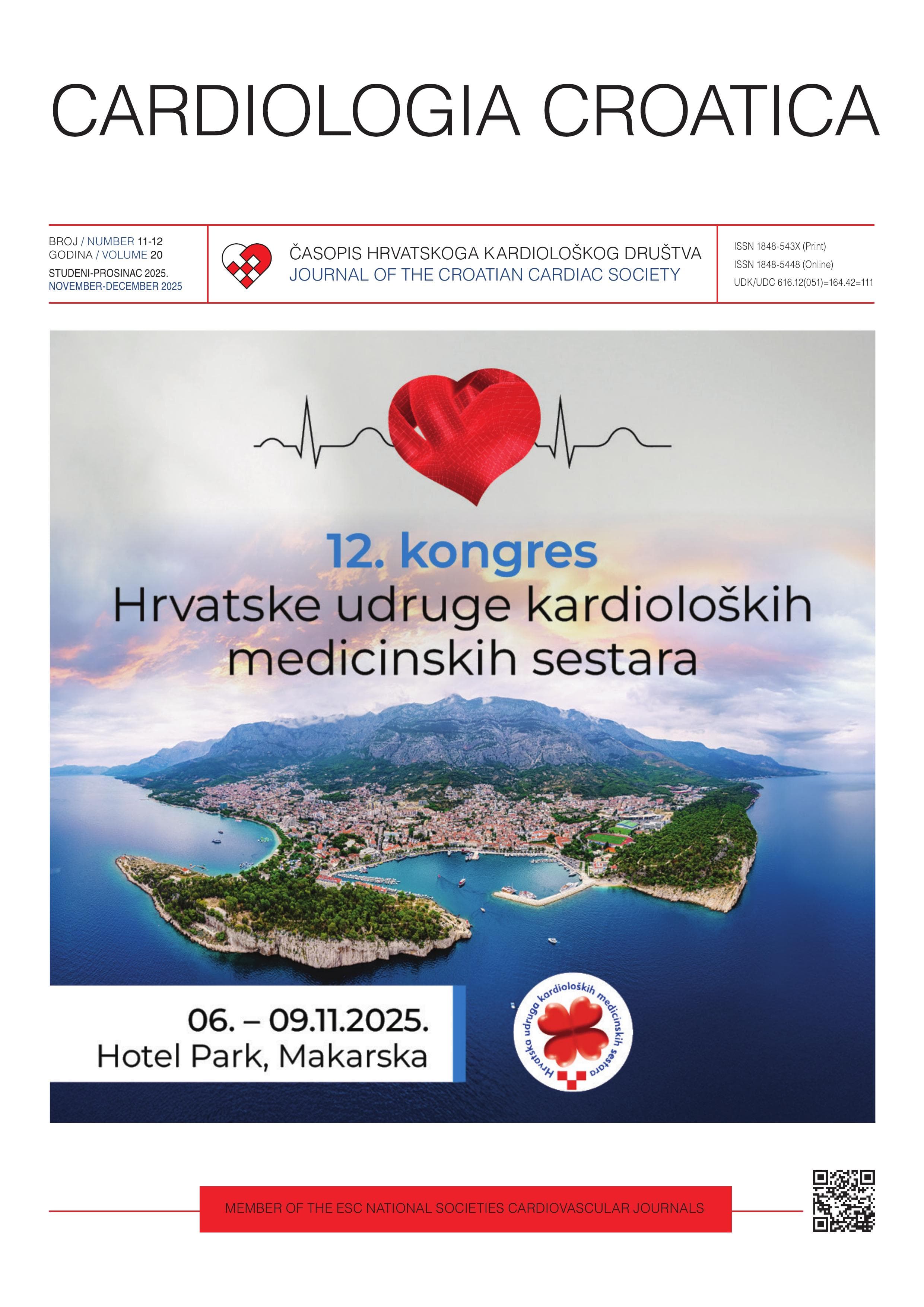
## Dear Colleagues, It is a great pleasure and honor to welcome you to the 12th Congress of the Croatian Association of Cardiology Nurses that will be held between November 6th and November 9th, 2025, at the Park Hotel in Makarska, Croatia. The Congress program is intended for medical nurses and related professions and represents an excellent opportunity for networking and sharing knowledge among all healthcare and non-healthcare professionals who are involved in the care for cardiovascular patients. The program will consist of plenary lectures, a conference of the working groups of the Croatian Association of Cardiology Nurses, sponsored conferences, workshops, and poster presentations. Your constructive discussions at these lectures, led by Croatian and international experts, will undoubtably spark the development of many new ideas. The numerous workshops that will be held as part of our Congress will also provide it with a practical educational dimension, helping participants acquire new or develop existing specific skills. Informal friendly discussions will provide participants with the opportunity to discuss any ideas or dilemmas they may have, so that we can all contribute to achieving the goal of our Congress together – fostering professional excellence in our field. We are all aware that advancements in biomedical and technological sciences introduce new challenges into cardiological practice at a rapid pace. We are proud to say that cardiological nursing in Croatia has kept up with the most recent trends and developments in cardiological practice, and the integration of new insights has remained focused on the individual and on preserving the fundamental values of medical care. Therefore, the focus and motto of this congress, just like the previous one, is “Person-centered care: mission, vision, and values”. At all our previous professional conferences, lectures on the everyday clinical practice of our participants were considered to be especially interesting. Our program therefore includes sessions describing interesting cases, brief reports from the everyday clinical practice of our participants, and poster presentations. Our participants will present their achievements, and you are all invited actively participate in the resulting discussions in order to exchange experiences, answer questions, and resolve any potential dilemmas. We would like to thank Prof Mario Ivanuša for his extensive assistance in the preparation and publication of congress abstracts in a supplemental issue of the Cardiologia Croatica journal. We are certain that our professional and friendly meeting in Makarska will remain a lasting and fond memory for all our participants. ## PREDSJEDNICI KONGRESA / CONGRESS PRESIDENTS: Ana Ljubas, mag. med. techn., FESC Ivica Benko, dipl. med. techn., ECDSAP
Ante Komazin, Gordana Hursa, Sanja Keleković, Tomislav Pijetlović
**Introduction**: Supraventricular tachycardias are among the most common symptomatic arrhythmias in childhood. Within this group, permanent junctional reciprocating tachycardia (PJRT) is rare but potentially life-threatening. PJRT typically presents in children and adolescents and is characterized by a concealed decremental accessory pathway with slow retrograde conduction. On electrocardiography, it manifests as a narrow QRS complex with a long RP interval and retrograde P waves. Unlike other supraventricular arrhythmias such as atrioventricular nodal reentrant tachycardia or atrioventricular reentrant tachycardia (AVRT), PJRT is often incessant and highly recurrent, carrying an increased risk of heart failure and tachycardiomyopathy. Clinical presentation is often nonspecific, ranging from palpitations and fatigue to reduced exercise tolerance and signs of cardiac decompensation. Definitive diagnosis relies on electrophysiological testing, while radiofrequency catheter ablation of the accessory pathway has emerged as a safe and highly effective treatment, providing definitive arrhythmia control and preventing long-term complications (1-3). **Case report**: We report the case of an 11-year-old girl who had been under cardiology follow-up since birth due to a muscular ventricular septal defect and intermittent Wolff-Parkinson-White syndrome. Her first symptomatic tachycardia episode occurred at the age of four, when ECG revealed intermittent atrial tachycardia with features of PJRT. Clinical examination and initial investigations excluded structural heart disease, while multiple ECG recordings confirmed the presence of supraventricular tachycardia. A 48-hour Holter monitor demonstrated persistent delta waves without paroxysmal supraventricular tachycardia episodes. Despite antiarrhythmic therapy, tachycardia persisted. Electrophysiological study confirmed the presence of both AVRT and PJRT, and radiofrequency ablation of two accessory pathways successfully terminated the arrhythmia. **Conclusion**: This case highlights the importance of timely recognition and treatment of PJRT in pediatric patients. Radiofrequency catheter ablation proved to be a safe and highly effective therapeutic option, preventing the development of secondary dilated cardiomyopathy or tachycardiomyopathy.
Luka Premužić
Cardiovascular disease remains the leading cause of death and disability in Croatia and worldwide, accounting for nearly one in every three deaths in Croatia and a significant share of healthcare expenditures. Coronary artery disease (CAD), the most common form, is frequently underdiagnosed or diagnosed only after acute events, leading to costly hospitalizations and avoidable mortality. MSCT Coronarography has emerged as a proven, non-invasive diagnostic tool with superior accuracy in detecting CAD compared to traditional stress testing. As a first of its kind in Croatia, the establishment of specialized MSCT Coronarography Unit in Dubrava University Hospital, made of nurses, X-ray technicians, cardiologists and radiologists represents a transformative step in modernizing cardiac diagnostics (1, 2). A dedicated specialized unit for CT coronarography can streamline care pathways, reduce invasive procedures, and lower system-wide costs by early, precise diagnosis, patient-centered care, cost efficiency and resource optimization, integration with preventive cardiology. With targeted government investment, robust implementation strategy, and Ministry of health collaboration, this initiative will reduce healthcare costs, save lives, and align Croatian cardiac care with global best practices.
Ivica Benko, Ivona Filipović, Magdalena Drljačić, Mateja Lovrić, Marina Žanić, Marina Budetić, Nikolina Slamek, Mirela Adamović, Marija Grlić
**Introduction**: Remote monitoring (RM) of cardiac implantable electronic devices shortens time to clinical decision from 22 to 4.6 days and reduces cardiovascular hospital stay from 4.0 to 3.3 days (1). RM can reduce in-hospital device evaluations by about 45% and detect clinically relevant events within less than 2 days (2). Network-based follow-up has been associated with improved survival compared with clinic-only care (3). We describe organizational challenges in introducing RM and early outcomes from a high-voltage (HV) device cohort. **Patients and Methods:** This was a single-center implementation integrating RM into routine workflow. A nurse-led coordination model was created with triage protocols, escalation pathways, electronic medical records templates, informed consent and General Data Protection Regulation-compliant data handling. The first 14 implantable cardioverter-defibrillator (ICD) / cardiac resynchronization therapy defibrillator patients were enrolled over four months. Alerts were reviewed on working days with predefined thresholds for intervention or recall. **Results:** Within the first four months, two device-related interventions were performed based on RM alerts and two patients were urgently recalled. One patient with heart failure presented with a new arrhythmia, and another experienced appropriate ICD therapy. No adverse events were linked to RM processes. Specially trained nurses managed alert handling and documentation with electrophysiology oversight. **Conclusion:** A structured nurse-led RM program for HV devices is feasible and safe, translating literature-proven benefits into clinical practice. Specially educated nurses can reliably manage daily RM operations, ensuring timely interventions and reduced outpatient load. Broader adoption requires formal education pathways, certification by professional societies and clear administrative and legal governance.
Verica Šeb, Mateja Šolić, Nikolina Glogovšek, Ivica Benko
**Introduction**: Critical illness neuropathy (CIN) is a diffuse peripheral neuropathy that occurs in patients with severe systemic illness, most frequently in sepsis or multi-organ dysfunction, and is associated with prolonged intensive care stay and mechanical ventilation (1). In heart transplant recipients, additional factors such as immunosuppressive therapy, recurrent infections, and metabolic disturbances may increase the risk and complicate recovery. **Case report**: We present a 61-year-old male patient with dilated and valvular cardiomyopathy who underwent orthotopic heart transplantation in December 2024. His postoperative course was complicated by infections (Klebsiella pneumoniae, Enterococcus faecalis, Candida glabrata), pleural and pericardial effusions, and recurrent cytopenias. During recovery, he developed progressive symmetrical muscle weakness and paraplegia. Neurological evaluation and clinical course were consistent with CIN. The patient required prolonged rehabilitation, nutritional optimization, and repeated hospital readmissions for endomyocardial biopsy and immunosuppressive adjustments. Despite complications, graft function remained stable, with ISHLT rejection grade 0–1R during follow-up biopsies. CIN in post-transplant patients poses unique diagnostic and therapeutic challenges, with no specific pharmacological therapy available. Management is primarily supportive, focusing on infection control, metabolic balance, early mobilization, and physiotherapy. Nurses play a pivotal role in recognizing early signs of neuromuscular weakness, preventing secondary complications (pressure ulcers, contractures, respiratory decline), and supporting rehabilitation. Nursing interventions in this case included assisted mobilization and transfer training, respiratory physiotherapy, glucose monitoring, and patient motivation in prolonged recovery. **Conclusion**: CIN remains an under-recognized but significant complication after heart transplantation. Early recognition, multidisciplinary collaboration, and targeted nursing interventions are essential to optimize functional recovery and quality of life in this vulnerable patient population.
Marina Radolović, Romina Tončinić, Maja Pištelek, Marica Komosar-Cvetković, Irena Kužet-Mioković
Migraines present a particular challenge in patients with cardiovascular diseases due to increased risk of heart attacks and strokes and the limited options for safe medications. Nurses play a vital role in assessing risk factors, educating patients, and supporting safe medication use. Epidemiological studies show that migraine, especially migraine with aura, increases cardiovascular risks (1, 2). Nurses should be aware that some migraine medications like triptans may not be safe for patients with cardiovascular disease (3). Newer preventive treatments such as CGRP receptor antagonists offer safer alternatives. Nurses should focus on teaching patients about lifestyle changes that reduce overall cardiovascular risk, including healthy diet, physical activity, stress management, and smoking cessation (4). Ensuring adherence to prescribed migraine therapy and monitoring for side effects are essential nursing responsibilities. A multidisciplinary team approach involving neurology, cardiology, and primary care, supported by nurse-led patient education and monitoring, is critical for optimizing care and reducing complications.
Andrijana Erak
In Anderson-Fabry disease (AFD), echocardiography is the first-line imaging modality and is crucial for prognostic stratification, clinical therapy, and screening. It is the rare X-linked genetic metabolic ailment known as AFD causes the enzyme α-galactosidase A to be deficient or absent, which causes glycosphingolipids to accumulate in several tissues and organs (skin, kidneys, blood vessels, central nervous system), including the heart. Increased myocardial inflammation, left ventricular hypertrophy, and myocardial fibrosis are the common outcomes of cardiac involvement. This is the most frequent cause of mortality for these people and can lead to a clinical spectrum of heart failure, restrictive cardiomyopathy, and arrhythmia episodes. When describing AFD cardiomyopathy, standard echocardiography is essential. Echocardiography is the best method for screening AFD cardiomyopathy since it is a diffuse, non-invasive, easily reproducible, and reasonably priced testing procedure (1, 2). In order to use echocardiography to screen for AFD, a nurse must secure the patient’s consent, educate them, prepare them for the procedure, provide supportive care, preserve their privacy, and assist with the practical aspects of the test. Additionally, echocardiogram examination is the most important imaging technique to help physicians follow up with patients who have AFD.
Antonela Barišić, Patricija Tomašković, Željka Božić, Martina Žderić
**Introduction:** Teamwork is one of the most important and challenging aspects of working in the healthcare system. Cardiological treatment of patients implies dynamism, speed, dexterity, skills and knowledge of healthcare professionals (1). The roles in the team chain should be adequately distributed, coordinated as a team, determined by time, and the size of the team must be optimal. Different cardiology departments and extensive treatment require the involvement of different experts, the functionality and good structure of the department, and adequate communication (2). **Methods:** The study was conducted in the University Hospital Centre „Sestre milosrdnice“ at the Clinic for Diseases of the Heart and Blood Vessels from March 30 to April 15, 2025. The study was attended by (N=79) nurses/technicians (85%), physiotherapists (7%), caregivers (4%) and radiology technicians (4%). The emphasis is on the nurse as a team leader. This study used a questionnaire consisting of 16 questions in two parts: functionality and leadership of the department and communication among respondents in the department. The questionnaire was translated and modified from the survey questionnaire – Team Stepps Teamwork Perceptions Questionnaire. **Results:** The results of research show that the majority of nurses/technicians are university-educated with the necessary experience and competencies. The function and leadership were evaluated satisfactorily in all segments; a certain part of respondents is less satisfied with the transfer of information by the manager or with each other and with the resolution of conflict situations. The results show satisfaction with teamwork, functionality and distribution of roles, emphasize the essentiality and expediency of the team. The survey shows that the team members agree with each other and support each other, correct each other, support and successfully resolve conflict situations. **Conclusion**: Teamwork is the most widespread form of work group when solving problems. When establishing a team, the goal, the size of the team, the division of roles and responsibilities and the time frame of the team duration are very important. (3) The goal must be the same for everyone, and everyone must strive to achieve that goal. The size and structure of the team must correspond to the complexity of the problem/goal that must be met. Roles and responsibilities are the most important aspects that must be adopted on the basis of character traits, abilities, experience, competence and the like (3).
Verica Šeb, Mateja Šolić, Nikolina Glogovšek, Ivica Benko
**Introduction.** Cardiovascular syphilis is a rare but serious late manifestation of Treponema pallidum infection, typically affecting the ascending aorta and coronary ostia (1, 2). It can mimic atherosclerotic disease and lead to aneurysm formation, aortic regurgitation, or ischemic heart disease (2, 3). Despite effective antimicrobial therapy, delayed or inadequate treatment may result in irreversible cardiovascular damage. **Case report.** We present a 71-year-old female with a history of arterial hypertension, dyslipidemia, chronic obstructive pulmonary disease, and prior ischemic stroke. During preoperative assessment in 2023, serology was positive for T. pallidum and she received benzathine penicillin G therapy. She was admitted for cardiological evaluation after incidental findings of an anteroseptolateral scar on 12-lead electrocardiography. Echocardiography showed mildly reduced left ventricular systolic function (LVEF 47%) with akinesis of the septum, apex, and basal inferior wall. Coronary angiography revealed chronic total occlusion of the left anterior descending (LAD) artery with collateral circulation, but without typical atherosclerotic features. Given her history of syphilis, cardiovascular tertiary syphilis was suspected. MSCT confirmed chronic LAD occlusion without aortic dilatation or signs of aortitis. Neurological, dermatological, and infectious disease consultations excluded neurosyphilis or cutaneous involvement. The patient remained hemodynamically stable during hospitalization and was discharged with optimized medical therapy and follow-up for myocardial viability testing. **Conclusion.** This case highlights the importance of considering tertiary syphilis in the differential diagnosis of atypical coronary artery disease. In addition to multidisciplinary collaboration, nurses play a pivotal role in patient care. Key nursing interventions included structured education about disease progression and medication adherence, as well as continuous monitoring for signs of heart failure (e.g., weight control, dyspnea assessment). These contributions not only support early detection of complications but also improve long-term outcomes and patient self-management.
Sebastian Sajko
Communication with a patient on mechanical ventilation is a significant challenge in intensive care units (1). It is one of the most important needs of the patient to exchange information and develop a relationship of trust (2). Due to the inability to communicate verbally, patients often feel a loss of identity, control and dignity. On the other hand, nurses and other staff in contact with the patient also experience frustration due to difficulty in understanding the patient’s wishes, attitudes and needs (2, 3). When effective and empathetic communication is established, patients feel safer, calmer and more actively involved in their own care and treatment. In practice, there are several ways and aids for communication. Lip reading, gesturing, writing on paper and communication boards are often not enough due to patient’s physical weakness, fatigue, tremors and other difficulties. Technological developments have enabled application of alternative and augmentative methods, such as devices that track eye movements and mobile applications. Applying these solutions can significantly reduce the feeling of isolation and increase the patient’s self-confidence. Successful communication requires, in addition to high and low technological solutions, a positive attitude, empathy, patience, presence, openness and the willingness of the staff to devote time and attention to the patients. Education about verbal and nonverbal communication and the use of available aids further contributes to the patient feeling understood and respected, thus improving the quality of care and emotional recovery. Communication with patients on mechanical ventilation should not be neglected. A combination of low and high-tech solutions, professional education, and an empathetic and patient approach ensures better cooperation, reduces stress, strengthens trust between patient and staff, and contributes to a faster patient recovery.
Iva Petković, Dubravka Milača
Explantation of a permanent pacemaker is a complex procedure that requires careful multidisciplinary planning to minimize complications and ensure patient safety. Indications for device removal include infection at the pocket site, systemic infection such as endocarditis, lead malfunction, device recall, or erosion of the device through the skin (1). Pre-procedural preparation involves thorough clinical and laboratory evaluation, optimization of anticoagulant and antiplatelet therapy, and exclusion of uncontrolled infections. Patients must be adequately informed and provide written consent, while preoperative measures typically include pre-procedural fasting, hair removal at the surgical site, and the establishment of reliable intravenous access. Strict aseptic conditions and perioperative antibiotic prophylaxis are essential to reduce the risk of reinfection. Depending on the complexity of the case, explantation may be performed via simple pocket revision, transvenous lead extraction, or surgical removal. Nursing care plays a key role in patient education, psychological support, intraoperative monitoring, and coordination of postoperative care, including wound management and follow-up. Standardized protocols based on international guidelines improve procedural success rates, reduce morbidity, and contribute to improved long-term patient outcomes.
Petar Horvat, Lucija Dizdarević, Saša Dizdarević
**Introduction**: Nursing care is based on a holistic approach, which involves a comprehensive view of the patient, not only through their illness but also through their physical, psychological, social, and spiritual needs (1). In patients with heart failure, this approach becomes especially important due to the complexity of the clinical picture, which is very often burdened with numerous comorbidities (diabetes, chronic obstructive pulmonary disease, hypertension). For this reason, nursing care requires individualized planning, flexibility in approach, and close cooperation with other members of the healthcare team. The nurse plays a key role in recognizing patient needs, implementing interventions, providing education, and offering support to both patients and their families. **Case report**: 67-year-old male patient was hospitalized at the Department of Cardiovascular Diseases, University Hospital Centre Osijek, with a diagnosis of heart failure. The patient presented to the Emergency Department with shortness of breath. He has a medical history of rheumatoid arthritis and chronic obstructive pulmonary disease. Symptoms had been present for five days prior, and the patient had initially consulted his general practitioner. Echocardiography showed an ejection fraction of 40% and severe aortic stenosis, while coronary angiography revealed normal coronary arteries. The patient received diuretic therapy and oxygen therapy. At the cardiac surgery consultation, an aortic valve replacement was indicated, which the patient initially refused. Despite the interventions provided, the patient reported no relief in symptoms. He expressed fear and inner restlessness. Through cooperation between nurses and physicians, the patient received appropriate education and nursing interventions to reduce fear, after which he agreed to undergo surgery. As a result of the interventions, the patient also learned new mechanisms for coping with fear and anxiety. On the fifth day of hospitalization, the patient developed oliguria. Blood tests showed worsening renal function, and on the sixth day, the patient underwent his first hemodialysis. Despite all therapeutic measures, renal function did not improve, and the patient began regular hemodialysis three times per week. Individualized and multidisciplinary nursing interventions significantly improve treatment outcomes in patients with heart failure and associated comorbidities. Such an approach leads to significantly better patient conditions, including symptom reduction, decreased rehospitalization rates, and improved psychological well-being. **Conclusion**: Providing adequate and high-quality nursing care to patients with heart failure and accompanying comorbidities requires detailed planning, teamwork among nurses, and involvement of other healthcare professionals. To facilitate and enhance the planning and implementation of such interventions, emphasis should be placed on the continuous education of current and future nurses in the field of care for patients with comorbidities.
Patricia Sigal, Andreja Virt, Dijana Tutić, Maja Marjanović
Although medical records date back to ancient Egypt, the systematic importance of nursing documentation began with Florence Nightingale, who recognized it as a foundation for nursing to evolve into a profession. In modern cardiology, patients are at risk of sudden clinical deterioration and require continuous monitoring, structured and accurate documentation is essential (1). We synthesized current evidence on the role of standardized nursing documentation in cardiology, with a focus on patient safety, continuity of care, and digital transformation. Recent studies demonstrate that standardized nursing documentation in cardiology improves early detection of clinical deterioration, enhances patient safety, ensures consistency of care, and reduces rehospitalization rates. The integration of digital technologies further advances documentation processes by enabling structured recording, facilitating interdisciplinary communication, and supporting decision-making. Amid current workforce shortages, standardization reduces errors and safeguards the registration of vital parameters. The three most important components of standardization today are: structured documentation templates, implementation of digital/electronic health records, and continuous staff education and training. Nursing documentation in cardiology serves not only as a communication tool but also as a clinical instrument directly influencing patient outcomes. The adoption of standardized forms, systematic training, and digitalization are key steps for improving quality of care in cardiac nursing practice.
Josipa Ribić
Peripheral arterial disease (PAD) is a chronic vascular condition that significantly impairs patients’ quality of life and increases the risk of cardiovascular complications. Its prevalence increases with age, particularly after the age of 45 (1). One of the typical symptoms is the so-called “walk in installments,” or intermittent claudication, which prevents patients from walking without interruption. Both symptomatic and asymptomatic PAD are indicators of myocardial infarction, stroke, and cardiovascular mortality (2). Identifying high-risk groups – smokers, patients with diabetes, hypertension, hyperlipidemia, and the elderly – is key to early disease detection. The educational poster for patients presents the main signs of the disease, preventive measures through lifestyle changes, and guidelines on whom to contact when symptoms occur. The crucial role of nurses in patient education and communication is especially emphasized, enabling earlier recognition of the disease, adherence to therapy, and reduction of complications.
Josipa Logožar, Ivana Babić
Certain congenital heart defects require surgical reconstruction of the right ventricular outflow tract (RVOT). Such anomalies, present in about 20% of children with congenital heart disease, include Tetralogy of Fallot, pulmonary atresia with or without ventricular septal defect, truncus arteriosus, transposition of the great arteries, and double outlet right ventricle (1). Over time, surgically implanted bioprostheses and valves undergo degenerative and calcific changes, which commonly lead to pulmonary stenosis or pulmonary regurgitation, and consequently to the need for reintervention (2). Until the year 2000, surgical replacement of the pulmonary valve was considered the gold standard of treatment. However, after the first successful transcatheter implantation performed by Bonhoeffer et al, pulmonary valve replacement, whenever indicated, began to be approached via the transcatheter or percutaneous route (2, 3). Currently, the three most commonly used valves are the Melody®, Edwards SAPIEN™, and Harmony® valves, with the Melody valve being used in Croatia. At the University Hospital Centre Zagreb, since 2019, when the first procedure was performed, 16 pulmonary valves have been implanted using the transcatheter approach, while more than 10,000 such procedures have been performed worldwide (2, 3). Before the procedure, the patient undergoes a comprehensive noninvasive and invasive cardiologic evaluation including electrocardiogram, echocardiography, chest X-ray, CT, MRI, coronary angiography, right heart catheterization, and laboratory blood tests. The procedure itself requires teamwork involving interventional cardiologists, anesthesiologists, radiology technicians, and nurses. The nurse’s role includes psychological and physical preparation of the patient, preparation of instruments and equipment, monitoring of the patient during and after implantation, recognizing changes in the patient’s condition, and promptly informing the physician. Transcatheter pulmonary valve replacement represents a significant advancement in the treatment of patients with congenital heart disease and RVOT dysfunction. Its minimally invasive nature, reduced risk of complications, and faster recovery emphasize the importance of expertise, skill, precision, and empathy among team members. The nurse’s knowledge and competencies in peri-procedural and post-procedural care are crucial for ensuring patient safety and a successful recovery.
Anita Pleško, Zrinka Paić, Julija Buljan, Josipa Pekez, Ivica Benko
**Introduction**: Cardiac tamponade is a rare but life-threatening complication of permanent pacemaker implantation, most commonly caused by lead perforation (1). Early recognition of the clinical triad and immediate response are crucial for survival. **Case report**: 74-year-old female was admitted following syncope. Medical history included arterial hypertension, moderate aortic stenosis, heart failure with preserved ejection fraction and coronary artery disease with prior percutaneous coronary intervention of the left anterior descending artery. Telemetry revealed a 6-second asystolic pause, and a dual-chamber permanent pacemaker (Biotronik Enitra 6 DR) was implanted. Several hours later on the ward, nursing staff observed sudden hypotension (BP 70/50 mmHg), atrial fibrillation (130/min) and signs of hemodynamic compromise. Bedside echocardiography confirmed a pericardial effusion up to 2.9 cm with right heart collapse, consistent with tamponade. Immediate fluid resuscitation and urgent triage ensured stabilization and rapid transport to the invasive laboratory. Pericardiocentesis via subxiphoid approach drained 220 ml of hemopericardium with prompt hemodynamic recovery (BP 120/80 mmHg, paced rhythm 60/min). The remainder of hospitalization was uneventful, and the patient was discharged in stable condition with recommendations for rehabilitation and regular device follow-up. **Conclusion**: Cardiac tamponade after pacemaker implantation is an uncommon but critical complication. In this case, rapid recognition of deterioration by nursing staff on the ward, initiation of volume resuscitation and activation of the transport pathway enabled timely pericardiocentesis. Rapid triage of symptoms and coordinated nurse-led action on the ward remain vital for patient safety.
Josipa Pekez, Zrinka Paić, Julija Buljan, Nikolina Valjak, Anita Pleško, Ivica Benko
**Introduction**: Ventricular tachycardia (VT) storm represents one of the most challenging entities in invasive arrhythmology. In patients with advanced ischemic cardiomyopathy and multiple ablation attempts, refractory VT can necessitate advanced heart failure therapies including transplantation (1). **Case report**: 65-year-old male with ischemic cardiomyopathy, history of myocardial infarction, multiple coronary interventions, and ICD implantation was admitted with sustained VT. Despite repeated catheter ablations (2023, Feb 2025, May 2025), recurrent polymorphic VT persisted. During the last procedure, multiple inducible VTs were documented, including unstable morphologies resistant to ablation. Hemodynamic compromise required advanced support and multidisciplinary discussion. Right heart catheterization confirmed elevated filling pressures and reduced cardiac index (2.3 L/min/m2). Owing to recurrent VT storm, severely reduced LV function (EF ~30%), and aneurysmatic remodeling, the patient was referred for urgent transplant evaluation. Comprehensive pre-transplant screening (dental, dermatological, urological, infectious, psychiatric) revealed no contraindications. On 13 June 2025, urgent orthotopic heart transplantation with ICD extraction was performed. The postoperative course was uneventful; the patient was weaned early from ventilation, mobilized within days, and later discharged on triple immunosuppressive therapy. Follow-up myocardial biopsies demonstrated no significant rejection (ISHLT 0–1R). **Conclusion**: This case illustrates the complexity of VT storm in end-stage ischemic cardiomyopathy and the ultimate role of heart transplantation. Successful management required coordinated work of arrhythmia specialists, heart failure cardiologists, intensivists, cardiac surgeons, and highly skilled nursing teams across cardiology and cardiac surgery. Nurses were integral in arrhythmia monitoring, advanced device management, perioperative care, and post-transplant education, highlighting the multidimensional role of nursing in one of the most complex areas of contemporary cardiology.
Ana-Maria Goronić, Mateja Lovrić, Marina Žanić, Marina Budetić, Nikolina Slamek, Mirela Adamović, Marija Grlić, Mario Tomašević, Ivan Horvat, Ivica Benko
**Introduction**: Generator replacement represents more than 25% of all cardiac implantable electronic device (CIED) procedures and carries a substantially increased risk of infection compared with de novo implantations (up to 2–4 times higher) (1). A randomized clinical trial in JAMA Cardiology including 427 patients demonstrated that the use of iodinated adhesive drapes reduced bacterial contamination from 9.9% to 2.9%, corresponding to a relative risk reduction of nearly 70% (2). This finding emphasizes a simple, evidence-based modification with direct implications for patient safety. **Results**: Immediately after publication, our center introduced iodinated adhesive drapes in all pacemaker and defibrillator replacements. The adoption was nurse-led, integrated into the existing workflow without prolonging patient preparation, and involved negligible additional cost. In the first two months after introduction, adherence was 100%, with seamless incorporation into sterile field preparation and no negative impact on procedural logistics. Nursing staff ensured protocol standardization, education, and real-time monitoring of compliance. **Conclusion**: The introduction of iodinated adhesive drapes for CIED replacements demonstrates how rapid translation of evidence into practice can significantly improve patient safety. Beyond infection prevention, this change highlights the importance of developing advanced nursing competencies, accepting new technologies, and continuously enhancing standards of care.
Irena Kužet Mioković, Marica Komosar-Cvetković, Kristina Skroče, Maja Pištelek, Dijana Travica Samsa, Ana Brajdić, Viktor Ivaniš, Marina Njegovan
**Introduction**: Cardiac rehabilitation (CR) is a cornerstone intervention for patients with cardiovascular and metabolic disease. Exercise-based CR programmes improve functional capacity, metabolic regulation, and psychosocial wellbeing, yet evidence from short-term interventions in patients with type 2 diabetes remains limited (1). **Methods:** Twelve patients with type 2 diabetes (age 65 ± 6 years, height 177 ± 12 cm, weight 89 ± 16 kg) participated in a 21-day pilot CR programme. Training included aerobic exercise, high-intensity interval training (HIIT), and resistance training, all individually tailored based on cardiopulmonary exercise testing (CPET) to define optimal training zones. **Results:** VO2 at VT2 increased from approximately 13.5 ± 3.0 to 15.5 ± 2.0 mL/min/kg after 21 days (p 2max showed an even greater improvement, rising from about 16.0 ± 2.5 to 18.0 ± 2.0 mL/min/kg (p < 0.001; d = 1.93), representing a very large effect size. Glycemic control improved, with time in range (3.9–10.0 mmol/L) rising from 87 ± 10% to 93 ± 5% by Week 2 (p < 0.05; d = 0.87) and remaining stable in Week 3. Time below range (<3.0 mmol/L) decreased from 2.0% to <1.0% (p < 0.05) and was nearly eliminated by Week 3 (p < 0.01). Time above range (10.0–13.1 mmol/L) fell from 9.1% to 5.2% (p < 0.05). SF-36 scores showed notable improvements in emotional limitations, physical health limitations, social functioning and emotional wellbeing. Pain remained unchanged, while physical functioning. **Conclusion:** A 21-day comprehensive CR programme combining aerobic, HIIT, and resistance training improved aerobic capacity, glycemic stability, cardiac stress biomarkers, and several quality-of-life domains in patients with type 2 diabetes. These findings highlight the multidimensional benefits of short-term CR, warranting confirmation in larger trials.
Barica Stanić, Željka Stojkov, Ivana Simić
**Introduction**: Cardiovascular diseases are one of the leading causes of death in the world. A key cause of it is acute myocardial infarction (AMI) which is caused by thrombosis of coronary arteries. It’s clinical picture commonly shows malign cardiac arrhythmia which can lead to cardiac arrest. Cardiac arrest is the most serious complication of AMI, with a high mortality rate and common lasting physical, cognitive and mental consequences (1). Immediate initiation of resuscitation, followed by cardiological stabilization, and subsequently comprehensive rehabilitation in which a multidisciplinary medical team and the patient’s family play an important role in survival and recovery from it. **Case report**: Patient K.I., born in 1993, was hospitalized after successful resuscitation at home. Family members initiated cardiopulmonary resuscitation until the arrival of Emergency Medical Services. Upon arrival, the emergency team registered ventricular fibrillation (VF). The patient was defibrillated four times before arriving at the hospital. In the hospital, he was endotracheally intubated, placed on mechanical ventilation, and underwent percutaneous coronary intervention (PCI) with stent implantation. After the procedure, VF recurred, requiring additional resuscitation measures. Repeat coronary angiography revealed marginal thrombosis in the stent, which was further dilated. The patient was admitted to the Intensive Care Unit. After three days, he was extubated and weaned from the ventilator. During the further course of hospitalization, the patient remained hemodynamically stable and was transferred to the Cardiology Department. The patient exhibited symptoms of depression, confusion, impaired communication, and non-cooperation. The family was allowed to stay with the patient throughout the day. With his wife’s assistance, the patient learned to speak, count, and form sentences. The nurse provided continuous health care, administered prescribed therapy, and offered support to both the patient and his family. The rehabilitation process involved a neurologist, psychologist, speech therapist, and physiotherapist. Good functional and neurological recovery was achieved, with preserved cardiac stability. After thirty days of hospitalization, the patient was transferred to a specialized neurological rehabilitation facility. **Conclusion**: The recovery of a patient after cardiac arrest is particularly complex and requires dedicated effort from medical staff. Active family involvement and consistent emotional support play a significant role in the patient’s psychological wellbeing and recovery.
Martina Vidak, Biljana Hržić, Ivica Benko, Valentina Miler
**Introduction**: Out-of-hospital cardiac arrest (OHCA) is commonly associated with metabolic and electrolyte disturbances, including acidosis, glucose dysregulation, and changes in potassium, calcium, magnesium, and lactate levels (1). Persistent abnormalities after return of spontaneous circulation (ROSC) are frequently linked with adverse outcomes. The aim of this study was to evaluate the prevalence of electrolyte imbalance and its association with survival in OHCA patients. **Results:** We retrospectively analyzed 38 patients with OHCA, 78% male, most frequently aged 70–79 years. The most common initial rhythm was ventricular fibrillation (79%). Potassium levels were normal in 53% of patients, hypokalemia was observed in 36%, and hyperkalemia in 7.9%. Regarding fluid therapy, 500 ml of normal saline was administered in 28.9% of cases, while the remainder received various combined infusions. Statistical analysis showed a significant difference in potassium levels between survivors and non-survivors (p=0.018). No statistically significant differences were observed for sodium, chloride, bicarbonates, or lactates. **Conclusion:** These findings emphasize the prognostic importance of potassium status in OHCA. Early recognition and correction of hyperkalemia, hypokalemia, or severe acidosis are crucial for resuscitation outcomes and survival, given the central role of electrolytes in cardiac function and conduction. Beyond clinical management, strengthening public education and first aid training remains essential, since a single trained bystander can determine survival. Clinical guidelines should therefore integrate both medical and preventive perspectives.
Matea Nikić, Ksenija Kasap, Ivica Benko
Early rehabilitation following pacemaker (PM) and implantable cardioverter-defibrillator (ICD) implantation represents a crucial step in the treatment of patients with arrhythmias and conduction disorders. While device therapy improves quality of life and survival, it carries procedural risks and require structured recovery strategies. Rehabilitation encompasses not only the physical restoration of function but also patient education and psychosocial adaptation to life with an implanted device (1). In the early postoperative period, emphasis is placed on vital signs monitoring, wound care, and prevention of complications such as bleeding, infection, or lead dislodgement. At the same time, maintaining upper limb mobility and preventing shoulder stiffness are achieved through light, supervised exercises. A gradual increase in range of motion and physical activity ensures safe reintegration into daily and occupational activities. Beyond physical recovery, structured education is essential. Many ICD patients experience anxiety related to potential shocks, which may limit activity and social participation. A multidisciplinary approach, involving cardiologists, nurses, and physiotherapists, provides comprehensive support to optimize outcomes. Early rehabilitation after PM and ICD implantation reduces complications, preserves functional capacity, and improves quality of life. Physiotherapists play a pivotal role not only in guiding safe physical activity but also in educating patients for long-term adherence to active lifestyles.
Igor Kafadar, Luka Zoričić
Acute conditions in intensive care medicine often require a rapid and adequate response from the entire medical team, including nurses and technicians. Sometimes, this means learning and applying methods that accelerate the diagnostic and treatment process for the patient. Ultrasound-guided fluid resuscitation is one of the techniques that nurses and technicians at the Department of Intensive Care Medicine at the University Hospital Centre Zagreb have successfully learned. When dealing with patients in acute hypovolemia, we encounter hypovolemic and distributive shock, where fluid administration is crucial for maintaining hemodynamic stability. By following the FALLS (Fluid Administration Limited by Lung Sonography) protocol for determining fluid resuscitation needs (1), physicians and nurses can simply and non-invasively assess the patient’s volume status, not relying solely on the display of the inferior vena cava, but also on pleural ultrasound, which shows the presence of A and B lines that clearly indicate pulmonary interstitial fluid overload. B lines, as hyperechogenic segments, clearly show the presence of solid material in the otherwise empty lung space and are a key indicator of circulatory overload or respiratory failure (2). In addition to the patient’s initial condition, lung ultrasound can also monitor the body’s response to fluid resuscitation and simultaneously rule out conditions that can disrupt this balance, such as cardiac tamponade and pulmonary embolism. The FALLS protocol has proven particularly useful in treating patients with weakened left ventricular function, as it allows for early detection and prevention of pulmonary edema, which can be fatal for the patient, while maintaining hemodynamics without excessive use of vasopressor support. The role of nurses and technicians in understanding ultrasound diagnostics and fluid resuscitation protocols can be crucial in life-threatening situations for patients. With their knowledge and skills, they will contribute to raising the standards of healthcare and intensive care medicine.
Lea Bago, Anita Martinović Vranješ
Cardiovascular diseases are the leading cause of morbidity and mortality worldwide, and early rehabilitation is crucial for reducing the risk of recurrent cardiovascular events and improving the quality of life of patient (1). The aim of this paper is to present the role of nurses in the rehabilitation process of patients with cardiovascular diseases, with a particular focus on cardiac rehabilitation. Rehabilitation programs are implemented through education, motivation, and continuous support. The paper provides an overview of the phases of cardiac rehabilitation, including the inpatient, subacute, and maintenance phases, while emphasizing the multidisciplinary approach in which nurses play a central role. It also examines the importance of physical activity, proper nutrition, adherence to therapy, and health literacy, alongside identifying barriers such as socioeconomic limitations, low motivation, and limited access to rehabilitation programs. Findings from the available literature confirm that nurses significantly contribute to cardiac rehabilitation, reducing complications and improving long-term outcomes for patients. It is concluded that the integrated and continuous role of the nurse is crucial for the successful implementation of cardiac rehabilitation and for the lasting improvement in the quality of life of patients with cardiovascular diseases (1).
Ivana Branilović, Monika Radić, Lucija Karamatić
MitraClip and TriClip are minimally invasive percutaneous procedures used to treat mitral and tricuspid regurgitation (1). MitraClip addresses severe mitral regurgitation, while TriClip targets tricuspid regurgitation. Both interventions reduce blood backflow through the affected valves and improve cardiac function. These procedures are particularly beneficial for patients who are not candidates for conventional surgery, offering reduced invasiveness, shorter recovery, and lower risk of complications (1). Preoperative care involves detailed medical history, laboratory and diagnostic tests, cardiac function assessment, and psychological preparation. Special attention is given to anesthesiologic evaluation and the placement of venous catheters. Postoperative care begins with intensive care unit monitoring, including vital signs, therapy administration, and observation for complications such as arrhythmia, bleeding, or infections. After stabilization, patients return to the cardiology ward for continued monitoring of valve function, intervention site care, and therapy. Early mobilization is initiated within 24 hours, and outpatient follow-up and therapy continue after discharge. These minimally invasive procedures significantly improve patients’ quality of life, reduce symptoms, prolong survival, and represent a modern, safe, and effective approach to the treatment of valvular heart disease.
Antonija Baković, Anita Martinović Vranješ
Aortic stenosis (AS) is the most common acquired valvular disease. Its prevalence is increasing as a result of population aging. When aortic stenosis becomes severe and symptomatic, valve replacement is indicated. Surgical aortic valve replacement is still considered the gold standard, but over the past two decades, minimally invasive transcatheter aortic valve implantation (TAVI) has proven to be an effective alternative to surgery. In most cases, the TAVI procedure can be performed using a retrograde transfemoral approach under local anesthesia. However, if the peripheral arteries are not of sufficient diameter for valve implantation, alternative access routes may be used, such as transaxillary, transcaval, transcarotid, transapical or direct aortic access (1). Aim is to present the key aspects of the TAVI procedure, including patient preparation, types of prosthetic valves, and potential perioperative complications. Nurses play an essential role as a member of TAVI team, either as scrub nurses or as those responsible for analgosedation and valve preparation. They also have a significant role in patient evaluation, preparation, and monitoring for complications. In alternative access approaches, the nurse’s role varies considerably depending on the type of access, primarily from an organizational standpoint, which makes these procedures more complex. Therefore, nurses must have a thorough understanding of alternative access routes so procedure preparation can be performed in the right way, as well as noticing and reacting to specific complications.
Hrvoje Topalović, Romana Ivelić, Ana Marinić
Cardiogenic shock is described as a clinical entity characterized by low cardiac output (less than or equal to 2.2 L/min/m2) resulting in organ hypoperfusion and tissue hypoxia (1). The most common cause is acute myocardial infarction, which leads to myocardial hypoperfusion and ischemia, as well as impaired left ventricular systolic and diastolic function and reduced myocardial contractility. The goal of treatment is to prevent irreversible damage to vital organs while restoring cardiac output. This is achieved by establishing a timely diagnosis, early initiation of drug therapy with respiratory support. Given the unfavorable prognosis and high mortality rate in cardiogenic shock, pharmacological therapy is often insufficient, and mechanical circulatory support is required to ensure adequate perfusion of vital organs. The first choice of circulatory support is the veno-arterial extracorporeal membrane oxygenation (VA-ECMO) system, which enables complete hemodynamic stabilization with simultaneous blood oxygenation, effectively ensuring systemic perfusion of vital organs. The use of the VA ECMO system leads to a decrease in end-diastolic volume and pressure due to blood extraction from the circulation, but retrograde blood return to the aorta increases the filling pressures of the already remodeled left ventricle, with reduced contractility, resulting in blood stasis in the aortic root and left ventricle, leading to the so-called “ECMO lung” (2). By using the microaxial pump – Impella CP, we compensate for the shortcomings of the VA ECMO system by aspirating blood from the left ventricle, which the pump ejects proximally into the ascending aorta, ensuring anterograde flow while increasing coronary artery perfusion and reducing end-diastolic pressure and left ventricular volume (3). Timely and optimal use of combined mechanical circulatory support (ECPELLA) is crucial for improving the outcome of treatment of patients with cardiogenic shock, and is becoming a standard therapeutic option in the treatment of patients.
Maja Marjanović, Andreja Virt, Paula Kontek, Arijana Ježek, Izidor Kranjčec
**Introduction**: Ventricular septal defect (VSD) is the most common congenital heart defect but may also occur as a life-threatening complication of acute myocardial infarction (1). Large defects can lead to pulmonary hypertension, biventricular failure, and significant valvular regurgitation. Alongside somatic manifestations, patients frequently experience heightened anxiety that negatively impacts recovery. Nursing staff play a key role in early detection and alleviation of anxiety, contributing to improved patient outcomes. **Case report**: 60-year-old male was transferred to the Acute Heart Failure Intensive Care Unit with a diagnosis of post-infarction VSD verified by echocardiography (17-25 mm defect, left-to-right shunt Qp/Qs 3.7). The patient reported fear of hospitalization, uncertainty about treatment, irritability, crying episodes, tension, and insomnia. Nursing interventions focused on continuous emotional support, patient education regarding diagnostic and therapeutic procedures, and active encouragement to verbalize symptoms of anxiety. The nursing team collaborated closely with physicians and involved family members to reduce patient distress. The patient was subsequently referred to cardiac surgery where surgical patch repair of the VSD was successfully performed. At the time of transfer, the patient demonstrated improved coping, self-recognition, and verbalization of anxiety symptoms, with decreased frequency of episodes. **Conclusion**: Mental health is an essential component of holistic cardiac care. Nurses are in a unique position to identify anxiety early, provide targeted interventions, and coordinate multidisciplinary support. Their role is crucial in empowering patients with complex cardiac conditions such as post-infarction VSD to achieve better psychological adjustment and quality of life.