Cardiologia Croatica
Journal Research Assistant
Journal Research Assistant
Journal Research Assistant
Journal Research Assistant
Matija Marković, Ivica Premužić Meštrović, Tomislav Letilović
Cardiologists are familiar with valvular heart diseases but often they do not perceive heart valves as source of ventricular arrhythmias (VA). In patients with structural heart disease (SHD) VAs originate from a substrate in diseased myocardium, while in patients without SHD most common origins are outflow tracts (OT) (1). Premature ventricular complexes (PVC) from OT are either from subvalvular, valvular or supravalvular myocardium as there are myocardial extensions above the semilunar valves, these VAs are created predominantly by mechanism of triggered activity or enhanced automaticity (1, 2). Papillary muscles, mitral and tricuspid anulus, aortomitral continuity and other sites in a structurally normal heart can also be the origin VAs. There are general and specific ECG characteristics that can localize the origin of VAs: bundle branch block type, axis, QRS polarity in lead V6, QRS duration, precordial transition, maximal deflection index, and so forth, therefore mapping of the PVC starts in the region that is presumed by ECG characteristic (3). While different anti-arrhythmic drugs (AAD) aim at different mechanisms; calcium channel blocker can suppress triggered activity and beta-clocker can suppress automaticity, in general ablation is more effective in clinically meaningful reduction of PVCs compared to AAD (up to 95% vs. up to 25%) (4). Ablations are generally safe but complication rates of catheter ablation in aortic root are not negligible and include myocardial rupture and tamponade, stroke, valvular damage, and coronary artery occlusion; these complications can be lowered by usage of intracardiac ultrasound (ICE) (1, 4). When mapping arrhythmias from the valves, we should stop perceiving the heart structures or valves as isolated parts and acknowledge that regional anatomy of these structures is among the most complex of those encountered by cardiac electrophysiologists, e.g., aortic valve is directly related with both atria, the interatrial septum, the right ventricular outflow tract and pulmonary valve, and the aortomitral continuity. Ablation is highly effective in suppression of PVCs, it has low numbers of complications, but ablation in aortic region brings serious complications to the table, therefore shared decision making with the patient and usage of ICE is of upmost importance.
Sandra Makarović
Mitral regurgitation is a common but complex valvular heart disease. Severe mitral regurgitation can produce left ventricular remodeling and finally irreversible left ventricular dysfunction. Echocardiography allows the reliable identification of the presence, severity, etiology, and mechanisms as well as the pathologic lesions of mitral regurgitation. The following questions relating to echocardiography should be asked before patients undergo surgery: (1) Is the mitral regurgitation organic or functional? (2) When is the optimal time for surgery? (3) What is the etiology of mitral regurgitation? (4) Where is the lesion(s)? (5) Can the mitral valve be repaired? (6) Which surgical technique is most appropriate? Quantitative assessment of the severity of mitral regurgitation is essential for the optimal timing of surgery. Transesophageal echocardiography as well as three-dimensional echocardiography can help to identify the etiology, mechanisms, and pathologic lesions prior to possible mitral valve repair. The optimal timing of mitral valve surgery in patients with severe mitral regurgitation is crucial. In recent years, mitral valve repair has been shown to be better than mitral valve replacement for both short- and long-term outcome. Important issues such as reducing operative mortality, improving long-term survival, and improving the rate of mitral valve repair are crucial when considering mitral valve surgery. (1, 2)
Matias Trbušić, Nikola Bulj, Ozren Vinter, Ivo Darko Gabrić, Marko Boban, Diana Delić-Brkljačić
The development of echocardiographic methods and magnetic resonance for the evaluation of heart structure and function raises the question of the role of right-sided heart catheterization (RHC) or invasive hemodynamic evaluation (IHE) in valvular heart diseases. According to current guidelines, this method occupies a peripheral role and is reserved for situations where non-invasive testing is non-inclusive or discordant with clinical status (1). As pulmonary hypertension (PH) is one of the criteria for surgery in asymptomatic aortic stenosis and mitral regurgitation (MR), it is especially important to measure it accurately. However, non-invasive measurement of PH based on echocardiography has its limitations and the possibility of error and is particularly problematic in the presence of severe tricuspid regurgitation (TR) and reduced right ventricular (RV) function. A typical example where this is particularly important is persistent severe TR after mitral valve surgery leading to dilatation and dysfunction of the RV. Cardiac reoperation carries an increased risk, but it is considered in the absence of left-sided valve dysfunction, severe RV or left ventricular (LV) dysfunction and severe pulmonary vascular resistance/hypertension where RHC plays a key role (1). A particular problem for cardiac evaluation is multiple and mixed valvular heart disease, especially in the presence of some other heart pathology such as coronary heart disease, LV dysfunction or constriction (2). Exercise RHC is becoming increasingly popular in the diagnosis of heart failure with preserved systolic function, and now it is a question of its role in the diagnosis of valvular disease as well. The test has the potential to predict a worse outcome and thus for earlier surgery in asymptomatic patients e.g. with aortic stenosis or MR (3)**.** In conclusion, IHE is a useful and relatively harmless tool in the examination of valvular heart disease, but experience of cardiology teams is needed to get a true benefit of the method and to avoid misleading information. Gaining experience requires hard work perhaps even in cases where IHE is not fully indicated.
Boško Skorić
Right ventricular failure (RVF) refractory do medical therapy carries a high mortality risk, and circulatory support with right ventricular assist device (RVAD) may be a lifesaving option. (1-3) While surgical RVAD represents a more invasive therapeutical approach that carries about 50% mortality, the use of percutaneous RVAD support may improve outcomes. Acute RVF may result from 1) decreased contractility due to RV myocardial infarction or myocarditis; 2) volume overload, i.e. increase in preload due to excessive transfusions or following LVAD implantation; 3) pressure overload, i.e. increase in afterload during hypoxia, acidosis, or positive pressure ventilation. RV displays distinctive features in comparison to LV, including high complaint and low resistant pulmonary circulation. Accordingly, RV is more sensitive to an increase in afterload. Treatment of acute RVF includes specific causative treatment, optimization of preload, management of arrhythmia, reduction of afterload, and augmentation of contractility. If RVF is refractory, it is crucial to consider mechanical circulatory support on time. Percutaneous RV support includes VA-ECMO, Impella RP axial pump, and ProtekDuo cannula with an extracorporeal centrifugal pump. The selection of the device depends on whether RVF is isolated or part of biventricular failure, and whether it is a consequence of pulmonary artery (PA) disease. Percutaneous or surgically implanted RVAD is an option in isolated RV failure. Although ECMO can be also used in this setting, it is the device of choice if RVF is a consequence of PA disease. In the case of biventricular failure, a patient can be supported with paracorporeal surgically implanted or percutaneous BiVAD or ECMO. Support in RVF may be a bridge-to-recovery, bridge-to-candidacy, bridge-to-heart transplantation, or bridge-to-permanent RVAD. RECOVER RIGHT study showed 73% survival in patients supported with Impella RP. The Protek Duo cannula with internal jugular cannulation allows early ambulation during support. It is crucial to understand the pathophysiology of RVF, i.e. recognize ventricular interdependence with impaired LV filling to avoid futile therapeutical strategies like excessive volume infusion, and to start with mechanical support before irreversible multiorgan failure develops.
Petra Grubić-Rotkvić, Zrinka Planinić
Primary degenerative mitral regurgitation (MR) is caused by abnormality of one or more components of the valve apparatus, whereas functional MR is a left ventricular (LV) disease. Mitral valve (MV) surgery is required to address the primary process in degenerative MR (DMR), but the timing of MV surgery is controversial in asymptomatic patients. The “watchful waiting” strategy is dictated by a timely LV dysfunction recognition. The guidelines recommend MV surgery in asymptomatic patients with LV ejection fraction (EF) <60%, LV end-systolic diameter 40 to 45 mm, new-onset atrial fibrillation, resting systolic pulmonary artery pressure (sPAP) exceeding 50 mmHg, or if surgical risk is low with high likelihood of durable MV repair (1). Current accepted echocardiographic measurements of LV function are not sensitive enough for early myocardial injury detection since end-systolic dimensions and EF are affected by the different loading conditions in MR. The key is to identify hemodynamic consequences early in MR time course so that timely surgery may reverse them before becoming irreversible (2). Studies struggle on identifying other parameters or methods that could detect subclinical LV systolic dysfunction and predict postoperative outcomes such as: 1) brain natriuretic peptide (BNP) (there is no specific BNP cutoff of BNP for intervention, but a serial increase in BNP may reflect LV dysfunction and increased reliance on preload reserve in maintaining cardiac output); 2) LV global longitudinal strain (GLS) (a sensitive method for early detection of LV dysfunction, independently associated with mortality, add incremental data to surgical timing, with cutoffs −17.9% and −21.7% shown in studies that differentiate patients at higher risk, and with the possibility to detect relative change of GLS from baseline as in cardio-oncology); 3) left atrial size and strain; 4) echocardiographic exercise testing (good contractile reserve measured by LVEF and LV GLS and preserved exercise capacity without sPAP or MR worsening are reassuring parameters for a beneficial outcome); 5) cardiac magnetic resonance imaging (it can potentially reclassify MR severity providing valuable data on LV function and volumes) (1-3). Lastly, determination of proper timing for DMR surgery remains challenging and requires more studies.
Katica Cvitkušić Lukenda, Domagoj Vučić, Marijana Knežević Praveček, Krešimir Gabaldo, Domagoj Mišković, Blaženka Miškić, Ana Livun
Valve diseases have a large share in the total morbidity and mortality of the adult population, and can be congenital or acquired. The last few decades have seen a predominance of degenerative (calcified) heart valve diseases due to a prolonged life expectancy in economically developed countries. The therapeutic approach has remained unchanged and in the case of severe dysfunction, the valve is replaced with a mechanical or biological prosthesis, balloon valvuloplasty or valve reconstruction. The findings in understanding the development of heart valves along with human genome sequencing have led to the discovery of a genetic basis in valvular diseases (1). Also, there are numerous evidence to suggest that heart valve diseases which develop in adulthood has its source in embryonic development. In this review authors will display the genetic basis of the two most common inherited valvular diseases: bicuspid aortic valve and mitral valve prolapse, as well as a review of the findings suggesting a genetic contribution to calcified aortic valve disease (2). In addition, this review will include a review of the guidelines and benefits of genetic testing, as well as highlight the need to include genetic counseling in families with proven or suspected malformations. Linking genetic information to the clinical phenotype (**Table 1**) and potential outcomes of surgical treatment leads to a personalized approach to each patient (3). ### TABLE 1: Gene mutations associated with valvular heart disease. | | **Location** | **Gene** | **Inheritance** | **Phenotype** | | --- | --- | --- | --- | --- | | **Bicuspid aortic valve** | | | | | | **Syndromic** | 17q24.3 | *KCNJ2* | AD | Andersen syndrome | | **Nonsyndromic** | 9q34.3 20q13.33 15q22.31 11q24.2 | *NOTCH1* *GATA5* *SMAD6* *ROBO4* | AD AD, AR AD AD | AOVD1 CHTD5 AOVD2 AOVD3 | | **Mitral valve prolapse** | | | | | | **Syndromic** | 15q21.1 9q22.33 3p24.1 7q21.3, 12q13.1, 2q32.2,9q34.3 | *FBN1* *TGFBR1* *TGFBR2* *Collagen types I–III, V/ XI* | AD AD AD AR,AD AD,AD | Marfan syndrome Loeys-Dietz syndrome 1 Loeys-Dietz syndrome2 OI 1,Ehlers-Danlos syndrome, cardiac valvular type | | **Nonsyndromic** | Xq28 | *Filamin A* | XL | Cardiac valvular dysplasia | | **Aortic valve stenosis** | | | | | | **CAVD** | 9q34.3 | *NOTCH1* | AD | AOVD1 | [†] AD – autosomal dominant, AR – autosomal recessive, AOVD – aortic valve disease, CHTD – congenital heart disease, OI – osteogenesis imperfecta, XL – X-linked, CAVD - calcified aortic valve disease.
Ivana Jurin, Tomislav Šipić, Daniel Unić, Igor Rudež, Šime Manola, Irzal Hadžibegović
Transcatheter aortic valve implantation (TAVI) has rapidly evolved over the past decade in response to growing population of older adults with aortic stenosis (1). Initially, TAVI was approved for patients determined to be inoperable, but recently has expanded to those who were qualified as intermediate and low risk. The appropriate patient for a TAVI procedure typically has symptomatic senile degenerative AS of a trileaflet valve but recently indications for TAVI have expanded beyond typical and therefore choosing the optimal candidate has become more challenging. Patient selection for TAVI is based on accurate clinical and anatomical assessment of aortic stenosis. Patient selection is crucial when determining whether a patient is likely to benefit from catheter-based intervention as opposed to surgical intervention as well as determine whether patient is ill with the aortic stenosis or ill because of the aortic stenosis. This is because there are several clinical and anatomical aspects to be considered by the Heart team that favors a particular approach to treating severe aortic valve disease. The Heart team needs to discuss and evaluate expected quality of life versus risk of complications (2). Besides of proper patient selection, the safety and efficacy of prothesis implantation depends on procedural guidance which is based on a multimodality imaging approach. There are several patient factors and comorbidities which are associated with a poor post-TAVI outcome. These comorbidities include respiratory conditions associated with chronic lung disease, chronic kidney disease as well as frailty. Mobility, nutrition, and cognition in patients have been incorporated into many preoperative evaluations for selecting TAVI patients due to the relationship with poorer outcomes. Identifying the comorbidities which lead to poor outcomes post-TAVI remains a particularly challenging issue. Larger trials are required to determine which subpopulations of patients stand to benefit most from this procedure.
Dean Strinić, Tea Friščić, Jasna Čerkez Habek, Jozica Šikić
Bicuspid aortic valve (BAV) is the most common congenital heart disease with an estimated prevalence between 0.5% and 2% (1). BAV is usually made of 2 unequal-sized leaflets where the larger leaflet has a central raphe or ridge that results from fusion of the commissures. Most common is the fusion of the right and left cusps which is associated with coarctation of the aorta. Pure BAV is rare, and it has symmetrical leaflets or there is no raphe (2). The majority of BAV are an isolated birth defect, but there are also genetic causes. It can appear as a part of a syndrome (e.g., Shone’s syndrome, Turner syndrome, Williams syndrome, Anderson syndrome), or because of a single gene mutation such as NOTCH1 and ACTA2, or a combination of multiple gene mutations. Recently, the role of the extracellular matrix role in the differentiation of the cells and formation of the leaflets is being investigated (1, 2). Because 20-84% of patients with a BAV develop ascending aortic dilatation, BAV should be regarded as a valvulo-aortopathy. The dissection incidence in BAV patients is eight times higher than in the general population (3). The clinical presentation of patients with BAV varies from mild to severe valve disease and the symptoms typically develop in adulthood. The clinical manifestations depend on the function of the aortic valve, the aortopathy and acquired complications such as endocarditis. Infectious endocarditis affects 10-30% of BAV patients during their lifetime and is more common in younger male patients (1). Due to abnormal hemodynamics on the aortic wall and different activity of the matrix metalloproteinases, BAV is associated with dilatation in the aorta, especially the aortic root and ascending aorta, and coarctation of the aorta often occurs simultaneously. With the incidence between 59% and 81%, aortic valve stenosis is the most common complication of the BAV, with calcification playing a major role (1, 2). Treatment strategies vary depending on the severity of the disease and dilation of the aorta. Together with valve replacement, transcatheter aortic valve implantation is now considered as an alternative option with good results (1-3).
Nikola Bulj
According to the recent European Society of Cardiology (ESC) guidelines aortic valve (AV) repair is the preferred surgical treatment in younger patients with severe aortic regurgitation (AR) and/or proximal aorta aneurysm. However, this surgical option is still underused in clinical practice so close cooperation between dedicated and expert surgeons and echocardiographers is mandatory to achieve best results (1). Careful echocardiographic examination with 3D transesophageal echocardiography is needed to determine aortic root and cusp pathology as well as disease of proximal ascending aorta. This includes definition of the aortic phenotype, which relies on the measurement of the aortic annulus, the sinuses of Valsalva, the sinotubular junction and the ascending aorta. AV disease is based on the distinction between a bicuspid and a tricuspid valvular phenotype. The assessment of valve dysfunction is based on cusp motion and jet characteristics. There are three types of valve dysfunction: type I characterized by normal cusp motion, associated with root/ascending aorta dilatation (central jet), type II defined as cusp prolapse (eccentric jet) and type III characterized by cusp restriction, associated with poor tissue quality and or quantity. Additional measurements could favor successful repair: valve retraction (a geometric height <16 mm in a tricuspid AV or <19 mm in a bicuspid valve) contraindicates the repair; preoperative effective height <9 mm helps with the decision of cusp resuspension. Also, a coaptation length ≥5 mm post AV repair predicts a more durable result. The effective height is the orthogonal distance from the annulus to the middle of the free margin of the cusp and the geometric height, also known as the cusp height, is defined as the distance between the cusp nadir and the middle of the free margin. The coaptation height, also called the coaptation length), is defined as the distance of cusp apposition in diastole (2). Heart team cooperation can improve patient selection for aortic valve repair and post-repair durability of competent aortic valve.
Petra Grubić Rotkvić, Zrinka Planinić, Ines Zadro Kordić, Ognjen Čančarević, Tea Friščić, Ante Pašalić, Jozica Šikić
**Introduction**: Prosthetic valve endocarditis (PVE) represents 10-30% of all cases of endocarditis, occurring in 1 – 6% of patients with prosthetic valves. Diagnosis of PVE is made according to the modified Duke criteria that are less sensitive than for native valve endocarditis (NVE) given limitation of echocardiography (1). Even though 18F-fluorodeoxyglucose positron emission tomography/computed tomography (FDG-PET/CT) is a more accurate diagnostic tool for PVE included as a major criterion in the European Society of Cardiology (ESC) Guidelines for the management of infective endocarditis (IE), increased periprosthetic FDG uptake not always reflects infection (2). **Case report:** 75-year-old male patient with a prior history of Bentall procedure in 2013 has been hospitalized for fever of unknown origin. Blood cultures came positive on Streptococcus pluranimalium and a high suspicion of PVE was raised. Transesophageal echocardiography (TEE) showed a suspected vegetation on mechanical aortic valve without compromising valve function (**Figure 1**). No other signs of septic embolization were found, except subconjunctival hemmorrhage. Since two major and two minor Duke criteria for definite IE were met, the patient was started on standard antibiotic treatment according to ESC guidelines. Despite completed targeted antibiotic treatment, the patient remained subfebrile without clear clinical manifestation of infection with repeated negative blood cultures and no signs of vegetation on TEE. FDG PET/CT was performed and showed diffuse perivalvular FDG uptake pattern consistent with inflammation. While awaiting for the PET/CT result, the patient became afebrile with no laboratory or clinical signs of infection. In consultation with cardiac surgeon a strategy of watchful waiting was taken. The patient remained stable during one year follow-up. Figure 1. Transesophageal echocardiography showing suspected vegetation on an mechanical aortic valve. **Conclusion**: Homogenous FDG uptake may be present in the perivalvular area in the absence of PVE, even years after valve implantation (1). Since the factors used to distinguish normal and abnormal FDG uptake patterns are not yet standardized (2), clinical judgement and multidisciplinary treatment approach are still crucial in decision making.
Tomislav Šipić, Ivana Jurin, Daniel Unić, Igor Rudež, Šime Manola, Irzal Hadžibegović
The occurrence of paravalvular leak (PVL) has been a matter of concern since the beginning of transcatheter aortic valve implantation (TAVI) not only because it was quite often but also because more than moderate PVL was an independent predictor of mortality (1, 2). The prevalence of PVL remains above 3.4% according to more recent series although it has decreased over time mainly due to more experienced operators and better patient selection. However, the ability of current TAVI valves to limit PVR in the noncompliant, calcified annulus is a result of improved procedural planning and techniques. Due to specific features of PVL which are often multiple, eccentric, and irregular, echocardiographic imaging in detection and quantification of PVL is challenging. Several of the parameters that are generally used to grade native aortic regurgitation are difficult to apply to the context of PVL. Recently, quantitative assessment of regurgitation with aortography has emerged and been validated, with favorable reproducibility and accuracy. PVL-graded moderate severity or greater is often associated with inappropriate positioning of the TAVI valve stent position, irregular stent shape due to eccentric calcium or raphe, and/or free space between stent and native annulus due to valve under-sizing or under-expansion. The use of corrective procedures may be considered depending on the severity of PVL and the anticipated risk of complications associated with these procedures. It is worth mentioning that most PVLs remain clinically silent, but 1-3% of patients with PVL require treatment due to symptoms of heart failure, hemolysis, or both. PVL closure is a complex and technically demanding procedure with a limited clinical experience. To the best of our knowledge to this date, no recommended approach or clear guidelines were proposed considering this procedure. More systematic and trial data are needed to make more definitive conclusions about management strategies.
Irzal Hadžibegović, Ivana Jurin, Tomislav Šipić, Daniel Unić, Igor Rudež, Šime Manola
**Background**: Patients scheduled for transcatheter aortic valve implantation (TAVI) have advanced age, with estimated more than 30% of patients with an indication for oral anticoagulation therapy due to atrial fibrillation (AF). Dual antiplatelet therapy after TAVI has been historically considered as a standard approach, with current guidelines supporting the use of oral anticoagulation monotherapy with vitamin K antagonists (VKA) in patients requiring stroke protection in atrial fibrillation. (1) Direct oral anticoagulants (DOACs) are being currently investigated as monotherapy in patients with AF after TAVI, with conflicting results among different agents. **Patients and Methods**: We analyzed 151 consecutive patients who underwent TAVI procedure in our institution from 2013 to 2021. **Results**: There were 67 (44%) patients with AF (paroxysmal AF in 17 (25%) patients) that underwent TAVI procedure. Their median age was 80 years, 24% had diabetes mellitus, 30% had concomitant coronary artery disease, and their median CHA2DS2Vasc score was 5 (high thrombotic risk). Pre-procedural anticoagulation therapy was AVK in 34 (51%), DOACs in 18 (27%), and the remaining 22% of patients were taking ASA or clopidogrel. Early post-procedural anticoagulation therapy was AVK in 44 (67%), DOACs in 7 (12%), with antiplatelet therapy in 14 (21%) of patients. One patient with AF had post-procedural stroke, with no cases of post-procedural stroke among non-AF group. Their in-hospital mortality was 3.4%, in comparison to 2.7% in patients without AF. After 2017, all patients with AF were anticoagulated with AVK or DOAC after TAVI. After 2019, when full percutaneous approach was introduced, 7 patients were managed with single DOAC early after TAVI (5 with apixaban, and 2 with rivaroxaban) and had no peri-procedural ischemic or bleeding complications related to anticoagulation therapy. **Conclusion**: Patients with AF scheduled for TAVI have increased bleeding and thrombotic risk and require scrutinized tailoring of anticoagulation and other concomitant therapy. With fast-track transfemoral TAVI and full percutaneous approach, early continuation of a single DOAC in optimal dose adjusted to age, renal function and other comorbidities appears to be safe and effective and needs to be evaluated in a larger cohort of patients.
Jozica Šikić, Petar Bešlić
Since its introduction at the turn of the century, transcatheter aortic valve implantation (TAVI) has created a revolution in the treatment of patients with symptomatic aortic valve stenosis. In the early days of TAVI we used only two different devices with limited sizes and access options, but now we have a wide range of commercially available transcatheter aortic heart valves. These valves differ significantly in design, configuration, and mechanism. They may be balloon-expandable, self-expanding, or mechanically expanded, and bioprosthetic leaflets may be intra- or supra-annular. Frame cell size and configuration, frame height, size range, and the design and profile of the delivery system and access sheath all differ. (1) Although many patients are often successfully treated with any of variety of valves, yet there are nuances that may lead to one valve being more suitable than another in a specific patient and anatomic subgroups, like younger patients, those with degenerative surgical bioprostheses (valve-in-valve TAVI), bicuspid aortic valves, severe left ventricular outflow tract and annular calcification or coronary artery disease.
Frane Runjić, Andrija Matetić, Ivica Kristić, Nikola Crnčević
**Background**: Rapid pacing during balloon aortic valvuloplasty (BAV) or transcatheter aortic valve replacement (TAVR) is an important step allowing for proper valve position and effective implantation while avoiding valve pop-up or ventricular complications. There are different regimes for rapid pacing during BAV/TAVR, some of which will be presented here (1). **Materials and methods**: Literature database PubMed has been reviewed yielding a total of 49 research articles related to rapid pacing during BAV/TAVR in the period of 2009 to 2021. Collected articles were assessed and reappraised to extrapolate different pacing options. **Literature overview**: Rapid pacing during BAV/TAVR can be achieved by insertion of a temporary transvenous pacemaker, reprogramming the existing permanent pacemaker in situ, or over-the-wire (OTW) pacing (1) (**Figure 1**). The latter further includes different protocols. The most utilized OTW pacing protocol is based on the pacing circuit consisting of a positive pole attached to the wire in the left ventricle (isolated with the catheter), while a negative pole is attached to the femoral subcutaneous tissue over the needle. However, while the described protocol offers an attractive and simple set-up, it carries a risk of contact loss, inadequate pacing, or possibly insufficient post-implantation pacing (2). Therefore, we present our modification of the OTW pacing scheme which allows for a safer procedure and bail-out options if necessary (**Figure 1**). Contrary to the conventional OTW scheme, our protocol includes the attachment of a negative pole to a wire in the inferior vein cava (5 French femoral sheath). This allows for stable contact, adequate length of the negative electrode, and quick bail-out insertion of a temporary transvenous pacemaker if necessary. Figure 1. Illustrative overview of rapid pacing methods during balloon aortic valvuloplasty or transcatheter aortic valve replacement. **Conclusions**: Rapid pacing during BAV/TAVR is an important procedural step, and different methods have been described to achieve this. Modification of the existing protocol may lead to additional benefits and a more convenient procedure.
Nikola Crnčević, Andrija Matetić, Frane Runjić, Ivica Kristić
**Background and Aims:** Balloon aortic valvuloplasty (BAV) is usually used as a bridge to percutaneous or surgical aortic valve intervention. While BAV is traditionally performed via transfemoral approach, transradial BAV is a safe and feasible alternative (1, 2). We present a case of BAV performed via transradial access at the University Hospital Centre Split, which to our knowledge, is a first time this procedure was performed in Croatia. **Protocol presentation**: 86-year-old lady was hospitalized on the vascular surgery department with symptoms of critical limb ischemia. Upon preoperative examination a strong heart murmur was noticed, with ECG changes suggestive of left ventricle strain. An echo was performed revealing an ejection fraction of 30%, and a low flow - low gradient aortic stenosis (MPG 38 mm Hg, and Vmax 3.7 m/s). A CTA of the aorta revealed a chronic infrarenal dissection and an occlusion of the right iliac artery, basically disabling the classic femoral access. We decided to perform a balloon aortic valvuloplasty using radial access, as described in the SOFTLY-II trial (3). Right radial access was obtained using a 6F sheath and a contralateral radial artery was cannulated for pressure monitoring during the procedure and a 5F sheath was placed in the femoral vein. After aortic valve crossing, a 260cm wire (Medtronic CONFIDA) was placed in the left ventricle apex. At that point the 6F sheath was exchanged with an 8F sheath (**Figure 1**). A non-compliant 18x40mm (Bard Atlas Gold) balloon was used during rapid pacing over the wire at 180/min (positive electrode at the short wire placed in femoral vein and negative on the wire in the LV). Periprocedural analgosedation with propofol in the bolus-continuous infusion scheme was used during the rapid pacing. Figure 1. Vascular access planning and feasibility. **Conclusions**: Transradial BAV is a safe alternative to transfemoral BAV, especially in old and frail adults waiting for TAVR, while minimizing the bleeding risk, and femoral access complications.
Ivica Kristić, Andrija Matetić, Frane Runjić, Nikola Crnčević, Jakša Zanchi, Matjaž Bunc
**Background and Aims**: Vascular closure devices (VCDs) have emerged as a routine method for vascular management. Their evolution allowed for further progress and development of interventional cardiology irrespective of the increased frailty of the treated population (1). This short review aims to present available VCDs and describe the most utilized regimes in everyday practice. In addition, we aimed to present a VCDs protocol at the University Hospital Centre Split. **Materials and Methods**: A literature search in the PubMed database was conducted yielding a total of 2,380 research articles in the period of 1975 to 2021. Out of these, 246 articles were review articles. A significant positive trend in the number of published articles was observed across the years (**Figure 1**). Figure 1. Overview of the indexed publications focused on vascular closure devices over the years. **Results**: Different types of VCDs exist according to the closure mechanism. Suture-based VCDs include Abbott Perclose ProGlide and Abbott Prostar XL. Plug-based VCDs encompass Terumo Angio-Seal, Terumo FemoSeal, Cordis MynxGrip, Teleflex Manta, Vivasure Medical PerQSeal, and InSeal Medical InSeal (2). The mechanism of vascular closure by both suture- and plug-based VCDs is presented in **Figure 2**. Large bore access often requires a combination of the different types of VCDs, and their utilization is literally limitless, depending on the center preferences. Among different options, the most utilized vascular closure method at the University Hospital Centre Split during transcatheter aortic valve replacement (TAVR) combines Abbott Perclose ProGlide and Terumo Angio-Seal allowing for adequate closure and vascular management (3). After the TAVR finalization, the percutaneous sutures are tightened around an 8 French sheath, followed by the insertion of the Terumo Angio-Seal device according to standards. Figure 2. Overview and mechanism of action of different vascular closure devices. **Conclusions**: In conclusion, VCDs represent a modern ubiquitary method for vascular access management, enabling safer and comfortable procedures in a wider patient population. Research on VCDs has substantially increased in recent years. Utilized regimes depend on the access size and center preferences.
Andrija Matetić, Marija Romić, Nikola Crnčević, Ivica Kristić, Vjekoslav Tomulić, Frane Runjić
**Background and aims:** Periprocedural analgosedation during transcatheter aortic valve replacement (TAVR) has evolved significantly over the past decade. First procedures were done in general anaesthesia under the supervision of an anesthesiologist, while recent trends suggest periprocedural analgosedation performed by interventional cardiologists/TAVR operators (1). This method often includes different medications and protocols, depending on the site-specific preferences. Here we present a review of propofol effects along with the protocol utilized at the Catheterization Laboratory (Cath Lab) at the University Hospital Centre Split. **Review and protocol presentation:** Our primary used anaesthetic medication is propofol due to its beneficial pharmacologic properties including short action and titratable effects leading to effective sedation with amnesia, well-being and anti-emesis (2). In addition, it shows a good correlation with bispectral index (BIS) allowing for easier titration during longer procedures. However, due to its insufficient analgesia, we utilize high dose-volumes of local anaesthetic and optionally a systemic bolus-doses of fentanyl in selected individuals (**Figure 1**). To this date, this protocol provided a safe, effective and reproducible periprocedural analgosedation during TAVR. Importantly, this protocol was successfully performed by the Cath Lab Team, without the need for additional healthcare resources or staff allocation (3). To avoid large radiation to the staff, we have also adjusted the equipment (use of long intravenous lines; rotation of X-ray tube; monitor orientation) and modified the staff position (**Figure 2**). FIGURE 1. Illustrative overview of propofol plasma concentrations and bispecter index in relation to procedural phases (Split protocol). FIGURE 2. Schematic layout of the equipment/staff in the Cath Lab with regard to analgosedation. **Conclusions**: In conclusion, there are different regimens for periprocedural analgosedation. Periprocedural analgosedation with propofol in the bolus-continuous infusion scheme proved to be safe, effective and reproducible during TAVR. Interventional cardiologists and specialized Cath Lab team are trained to provide all aspects of management and care to these patients.
Marko Perčić, Zrinka Planinić, Ante Pašalić, Tea Friščić, Daria Ljubas Perčić, Edvard Galić, Jozica Šikić
**Introduction**: Transcatheter aortic valve implantation (TAVI) is now a common procedure to treat high-risk patients with severe aortic stenosis (1). The incidence of post-TAVI infective endocarditis is 3.25%, but it is associated with high mortality and complications. The most common complication is heart failure with a cumulative incidence of 37.1%. Appropriate measures should be taken to prevent infective endocarditis in post-TAVI patients including adequate antibiotics prophylaxis (2). **Case report:** We present a patient with history of implanted TAVI in December 2019, a condition after myocardial infarction and consequent secondary ischemic cardiomyopathy. In August 2020 he was hospitalized again at our Clinic because of acute heart failure and fever. Treatment was initiated according to guidelines for acute heart failure with non-invasive ventilation. Echocardiography was performed (EF 40%), and a severe mitral regurgitation was verified (in two jets, one between A3 and P3 segment, and the other between A2 and P2 segment, PISA 1.2 cm, EROA 0.7 cm2, MR RV 66 ml) with a visible vegetation on the anterior mitral cusp, on the atrial side, measuring 12x6 mm. Paravalvular “leak” of artificial aortic valve was visible, which reached the anterolateral papillary muscle with its jet, with PHT 423 ms. Also, minor abscess cavity beside artificial aortic valve was detected. S. Aureus was isolated from blood cultures, and linezolid was first introduced into the therapy, followed by vancomycin. Additionally, septic liver embolizations were radiologically verified. The patient’s condition gradually deteriorated with volume retention and oliguria, and small doses of dobutamine (5mcg/kg/min) were administered. Significant clinical improvement was obtained with abundant diuresis and improvement in renal function. Patient was emergently transferred to the cardiac surgery by appointment for the planned surgical treatment of mitral valve endocarditis and TAVI removal with SAVR. **Conclusion**: The mortality rate of infective endocarditis is 25%. There is a higher incidence of infective endocarditis in those with prosthetic heart valves and cardiac devices. In these cases, urgent surgical intervention is necessary.
Jasna Čerkez Habek, Dean Strinić, Zrinka Planinić, Jozica Šikić
**Introduction**: Calcific aortic stenosis (AS) is a prevalent and worrisome healthcare problem. Patients with symptomatic severe AS have an indication for aortic valve replacement. Indication for operation remains controversal in patients with isolate, asymptomatic severe AS. North American and European guidelines agree on a class I indication for operation in patients with a reduced left ventricular ejection fraction (<50%) but are inconsistent for patients with other disease or comorbid factors (1). The natural history of aortic stenosis should be better quantified to improve understanding of benefits and harms of intervention vs conservative treatment (2, 3). Aim: searching for latest knowledge about management of asymptomatic, severe AS and identification of highest risk patients that will have benefit from early intervention. **Materials and Metods:** The meta-analysis of literature search 2370 observational studies with total of 4075 patients comparing an early surgical treatment strategy with watchful waiting (1). The primary end point was all-cause death during long-term follow-up. Meta-analysis was performed on the association of prognostic indicators with the composite of death or aortic valve intervention found in multivariable models. **Conclusion**: The optimal management strategy for asymptomatic patients with severe aortic stenosis remains unclear. Data suggest that many patients with asymptomatic severe AS develop an indication for aortic valve intervention, and their deaths are mostly cardiac but not only sudden. Meta-analysis indicates that intervention was associated with a significant reduction in all-cause mortality during follow-up. Variables associated with mortality-related outcomes indicates that prognosis is significantly worse if global longitudinal strain or valvulo-arterial impedance is present even with a preserved LV function, if AS is more severe as measured by higher valve gradient and lower valve area, and if atherosclerotic risk factors, such as dyslipidemia or diabetes, are present.
Zrinka Planinić, Petra Grubić Rotkvić, Jasna Čerkez Habek, Marko Perčić, Petar Bešlić, Edvard Galić, Jozica Šikić
**Introduction**: Left ventricular non-compaction (LVNC) is a rare congenital cardiomyopathy characterized by excessive trabeculation and deep intertrabecular recesses most commonly affecting apical and mid-ventricular inferior and lateral segments. These patients are more prone to develop a typical triad of heart failure, thromboembolic events, and malignant arrhythmias. LVNC often coexists with other congenital or valvular heart diseases. Echocardiography is the standard imaging for the diagnosis of LVNC with several proposed criteria, mainly based on the ratio of non-compacted to compacted myocardial thickness (1, 2). **Case report:** 75-year-old woman with a prior history of arterial hypertension and atrial fibrillation was hospitalized due to acute heart failure. Physical examination revealed holodiastolic precordial murmur, bilateral pulmonary crackles, and peripheral edema. Echocardiography showed severe regurgitation of tricuspid aortic valve based on the prolapse of the non-coronary cusp and volume overload – induced eccentric left ventricular (LV) hypertrophy with reduced ejection fraction (EF) of 35%. Moreover, a hypertrabeculation with intertrabecular recesses were observed in the inferolateral and apical region of LV fulfilling the echocardiographic diagnostic criteria for LVNC (**Figure 1**). Coronary angiography showed no signs of coronary artery disease. The patient was started on standard heart failure therapy and was referred to cardiac surgery for aortic valve replacement (AVR). Postoperative echocardiographic assessment one year after AVR showed no improvement in LV systolic function despite normally functional bioprosthetic aortic valve and standard of care heart failure therapy. FIGURE 1. Four-chamber view showing ratio of the non-compacted and compacted myocardium. **Conclusion**: Symptomatic LVNC patients with LV systolic dysfunction generally have poorer prognosis (1). Since the potential of recovery of myocardial function is questionable in patients with preexisting myocardial disease (2), the postoperative outcome might not be satisfactory.
Vlado-Vlaho Ćubela, Maro Dragičević, Joško Bulum, Tajana Filipec Kanižaj, Helena Jerkić
**Introduction**: Severe aortic stenosis (AS) in patients with end-stage liver disease (ESLD) increases the morbidity and mortality of orthotopic liver transplantation (OLT). OLT is a surgical intervention with serious hemodynamic changes which can contribute to heart failure and cardiac death. Due to increased perioperative mortality, severe AS and ESLD are considered contraindications for OLT or aortic valve replacement (AVR) (1). Aortic balloon valvuloplasty (BAV) may be a reasonable therapeutic option to improve the hemodynamic status and enable OLT (2). **Case report:** The patient was a 43-year-old female who had an OLT in 2019 due to fulminant hepatitis B. AS was detected in 2018 and it progressed to severe in 2021. In 2021, another decompensation of cirrhosis of the liver graft occurred. The patient had Child-Pugh B graft cirrhosis which is according to the available data, contraindication for cardiac surgery (3). A multidisciplinary meeting was held and it was decided to do a BAV as a less invasive procedure before OLT. BAV was made with a satisfactory reduction of the transvalvular pressure gradient by 30% and with this procedure, the patient is hemodynamically in a more favorable position for OLT. **Discussion**: BAV was presented as a minimally invasive procedure with fewer complications to surgical replacement for AS in patient with ESLD. In practice, the procedure was proved to have a high percentage of complications and only a modest hemodynamic upgrade compared with AVR. Therefore, valvuloplasty should not be the first choice for AS. However, there are some indications for BAV such as a palliative treatment and in patients with AS and ESLD as a bridging procedure to OLT (1). This procedure enabled our patient to undergo surgery in a hemodynamically more favorable situation. It is also necessary to say that a multidisciplinary approach involving gastroenterologists, cardiologists, cardiac and abdominal surgeons, and anesthesiologists is the key in the treatment of such patients. In conclusion, in patients who are unsuitable for OLT because of AS, BAV, and then OLT may be a good approach that permits AVR posttransplantation. However further research is needed to demonstrate the advantages and disadvantages of this approach.
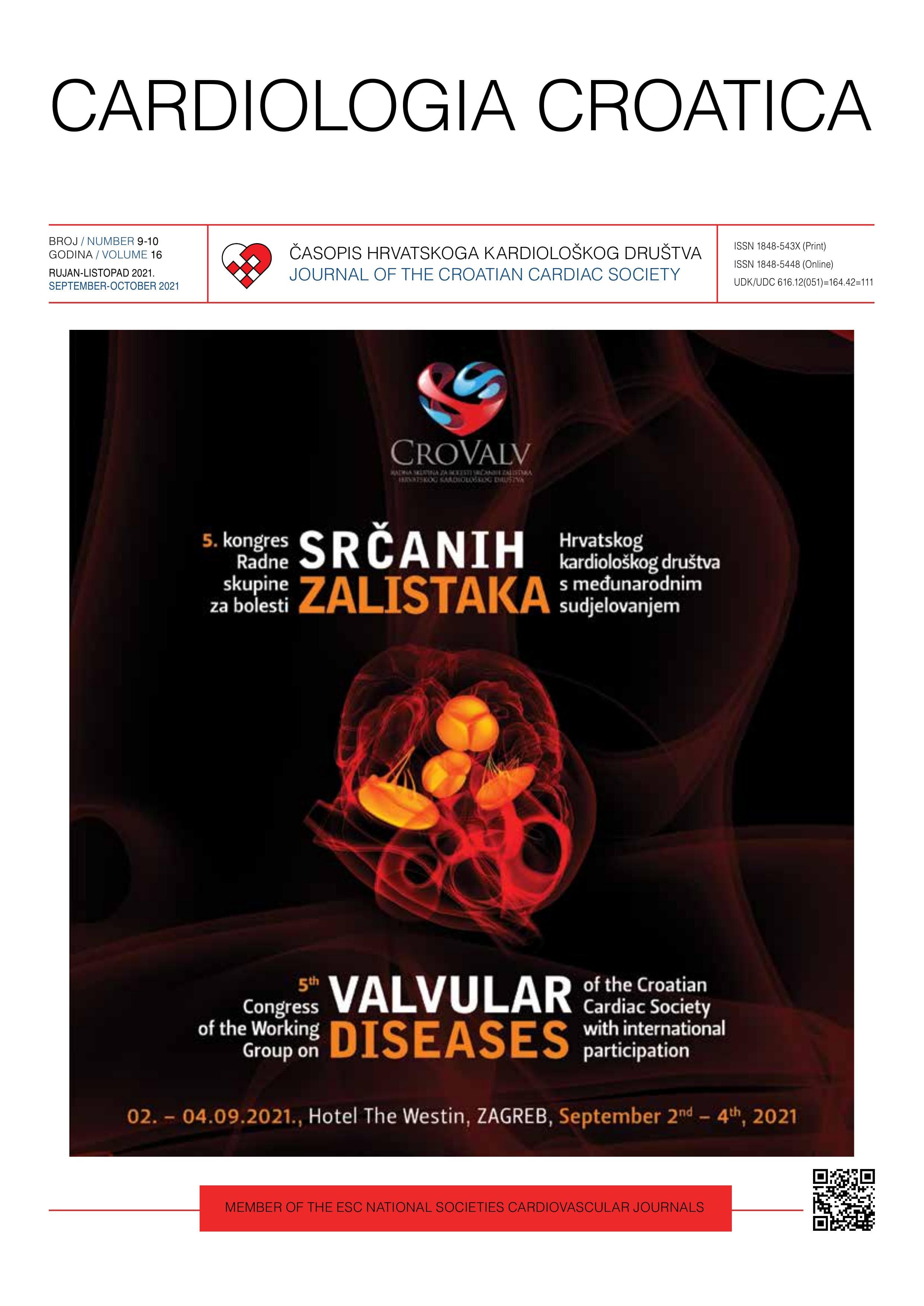
Jozica Šikić
The 5th Congress of the Working Group on Valvular Diseases of the Croatian Cardiac Society with international participation, CROVALV 2021, will be held on September 2nd to September 4th, 2021, at the Westin Hotel in Zagreb. CROVALV is a bi-annual Congress, designed for cardiologists, cardiac surgeons, anesthesiologists, neurologists, residents, general practitioners, and for all who are professionally involved in the treatment of valvular diseases. We have made a lot of progress since the last congress, especially in the field of percutaneous interventional treatment as well as in cardiac surgery, coronary artery diseases, and heart failure, which indirectly affects improvement of cardiac valve function. A lot of change has also happened in the field of preventive medicine and anticoagulation treatment. The goal of the congress is to present challenges in the approach, diagnosis, and selection of the most optimal way of treating patients with valvular diseases and to help implement the latest research findings into clinical practice. In addition to invited lectures by reputable speakers from abroad, leading national experts will bring their best. We hope that you will again recognize the value of the Congress and we cordially invite you to come and actively participate in contributing to the quality and success of the congress. Predsjednica kongresa / *Congress Director* doc. dr. sc. **Jozica Šikić**, dr. med. / *Assist Prof Jozica Šikić, MD, PhD* predsjednica Radne skupine za bolesti srčanih zalistaka Hrvatskoga kardiološkog društva